Comprehensive Clinical Periodontology Case Study Presentation
This comprehensive clinical periodontology case study presentation includes detailed information on the patient's general and dental history, oral hygiene assessment, periodontal diagnostics, radiographic analysis, occlusion evaluation, periodontal risk assessment, diagnosis, therapy plan, surgical therapy plan, and prognosis. The report also covers student information and includes dental photography, periodontal indices, gingival recession, keratinized gingiva width, tooth mobility, bone resorption, furcation involvement, and occlusion analysis.

Download Presentation

Please find below an Image/Link to download the presentation.
The content on the website is provided AS IS for your information and personal use only. It may not be sold, licensed, or shared on other websites without obtaining consent from the author.If you encounter any issues during the download, it is possible that the publisher has removed the file from their server.
You are allowed to download the files provided on this website for personal or commercial use, subject to the condition that they are used lawfully. All files are the property of their respective owners.
The content on the website is provided AS IS for your information and personal use only. It may not be sold, licensed, or shared on other websites without obtaining consent from the author.
E N D
Presentation Transcript
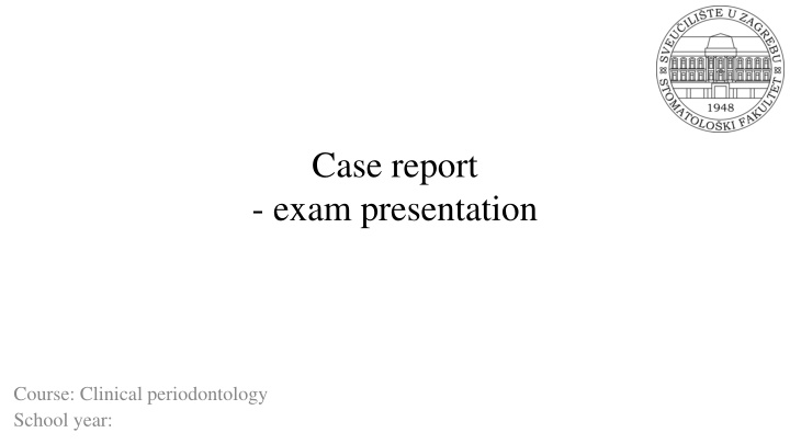
Case report - exam presentation Course: Clinical periodontology School year:
CONTENT BASIC INFORMATION HISTORY DENTAL PHOTOGRAPHY DIAGNOSTICS RADIOGRAPHIC ANALYSIS OCCLUSION ANALYSIS PERIODONTAL RISK ASSESSMENT DIAGNOSIS THERAPY PLAN SURGICAL THERAPY PLAN PROGNOSIS
STUDENT INFORMATION Name and surname Group School year GENERAL INFORMATION - PATIENT Chart number Initials Gender Year of birth
MEDICAL DENTAL HISTORY ORAL HYGIENE PERIODONTAL
DENTALPHOTOGRAPHY PHOTOGRAPHY FRONTAL PHOTOGRAPHY LATERAL PHOTOGRAPHY LATERAL
Aproximal plaque index(+ / ) API = V O 18 48 17 47 16 46 15 45 14 44 13 43 12 42 11 41 21 31 22 32 23 33 24 34 25 35 26 36 27 37 28 38 O V Papilla bleeding index (0 4) PBI = DIAGNOSTICS V O 18 48 17 47 16 46 15 45 14 44 13 43 12 42 11 41 21 31 22 32 23 33 24 34 25 35 26 36 27 37 28 38 O V Probing depth (mm) 18 48 17 47 16 46 15 45 14 44 13 43 12 42 11 41 21 31 22 32 23 33 24 34 25 35 26 36 27 37 28 38 Gingival recession (mm) V O 18 48 17 47 16 46 15 45 14 44 13 43 12 42 11 41 21 31 22 32 23 33 24 34 25 35 26 36 27 37 28 38 O V
DIAGNOSTICS Keratinized gingiva width (vestibular, mm) 18 48 17 47 16 46 15 45 14 44 13 43 12 42 11 41 21 31 22 32 23 33 24 34 25 35 26 36 27 37 28 38 Tooth mobility (0 3) 18 48 17 47 16 46 15 45 14 44 13 43 12 42 11 41 21 31 22 32 23 33 24 34 25 35 26 36 27 37 28 38
Bone resorption (1 3) 18 48 17 47 16 46 15 45 14 44 13 43 12 42 11 41 21 31 22 32 23 33 24 34 25 35 26 36 27 37 28 38 RADIOGRAPHIC ANALYSIS Furcation involvement (F1 F3) 18 48 17 47 16 46 15 45 14 44 13 43 12 42 11 41 21 31 22 32 23 33 24 34 25 35 26 36 27 37 28 38
Premature contacts OCCLUSION ANALYSIS 18 48 17 47 16 46 15 45 14 44 13 43 12 42 11 41 21 31 22 32 23 33 24 34 25 35 26 36 27 37 28 38 Protrusion contacts 18 48 17 47 16 46 15 45 14 44 13 43 12 42 11 41 21 31 22 32 23 33 24 34 25 35 26 36 27 37 28 38 Right laterotrusion - contacts 18 48 17 47 16 46 15 45 14 44 13 43 12 42 11 41 21 31 22 32 23 33 24 34 25 35 26 36 27 37 28 38 Left laterotrusion - contacts 18 48 17 47 16 46 15 45 14 44 13 43 12 42 11 41 21 31 22 32 23 33 24 34 25 35 26 36 27 37 28 38
Probing depth (mm) 18 48 17 47 16 46 15 45 14 44 13 43 12 42 11 41 21 31 22 32 23 33 24 34 25 35 26 36 27 37 28 38 PROGNOSIS Furcation involvement (F1 F3) 18 48 17 47 16 46 15 45 14 44 13 43 12 42 11 41 21 31 22 32 23 33 24 34 25 35 26 36 27 37 28 38
OPTIONAL: re-evaluation probing depths and dental photography BASELINE, BEFORE THERAPY AFTER THERAPY RE-EVALUATION Probing depth (mm) Probing depth (mm) 18 17 16 15 14 13 12 11 21 22 23 24 25 26 27 28 18 17 16 15 14 13 12 11 21 22 23 24 25 26 27 28 48 47 46 45 44 43 42 41 31 32 33 34 35 36 37 38 48 47 46 45 44 43 42 41 31 32 33 34 35 36 37 38 PHOTOGRAPHY PHOTOGRAPHY


")

")


")