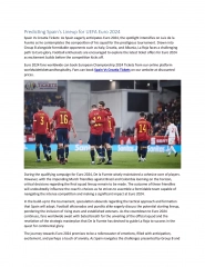
Comprehensive Guide to Paranasal Sinuses
Learn about paranasal sinuses, which are essential air-filled spaces within the skull and facial bones. Explore detailed information on the different types of paranasal sinuses, including the maxillary, frontal, sphenoidal, and ethmoidal sinuses. Discover the developmental aspects of the maxillary sinus and its anatomical characteristics. Gain insights into the growth of sinuses from birth to adulthood and their functional contributions to olfactory and respiratory physiology.

Download Presentation

Please find below an Image/Link to download the presentation.
The content on the website is provided AS IS for your information and personal use only. It may not be sold, licensed, or shared on other websites without obtaining consent from the author. If you encounter any issues during the download, it is possible that the publisher has removed the file from their server.
You are allowed to download the files provided on this website for personal or commercial use, subject to the condition that they are used lawfully. All files are the property of their respective owners.
The content on the website is provided AS IS for your information and personal use only. It may not be sold, licensed, or shared on other websites without obtaining consent from the author.
E N D
Presentation Transcript
Contents Introduction Typesof paranasal sinuses 1)Maxillary sinus 2)Frontal sinus 3)Sphenoidalsinus 4)Ethmoidal sinus
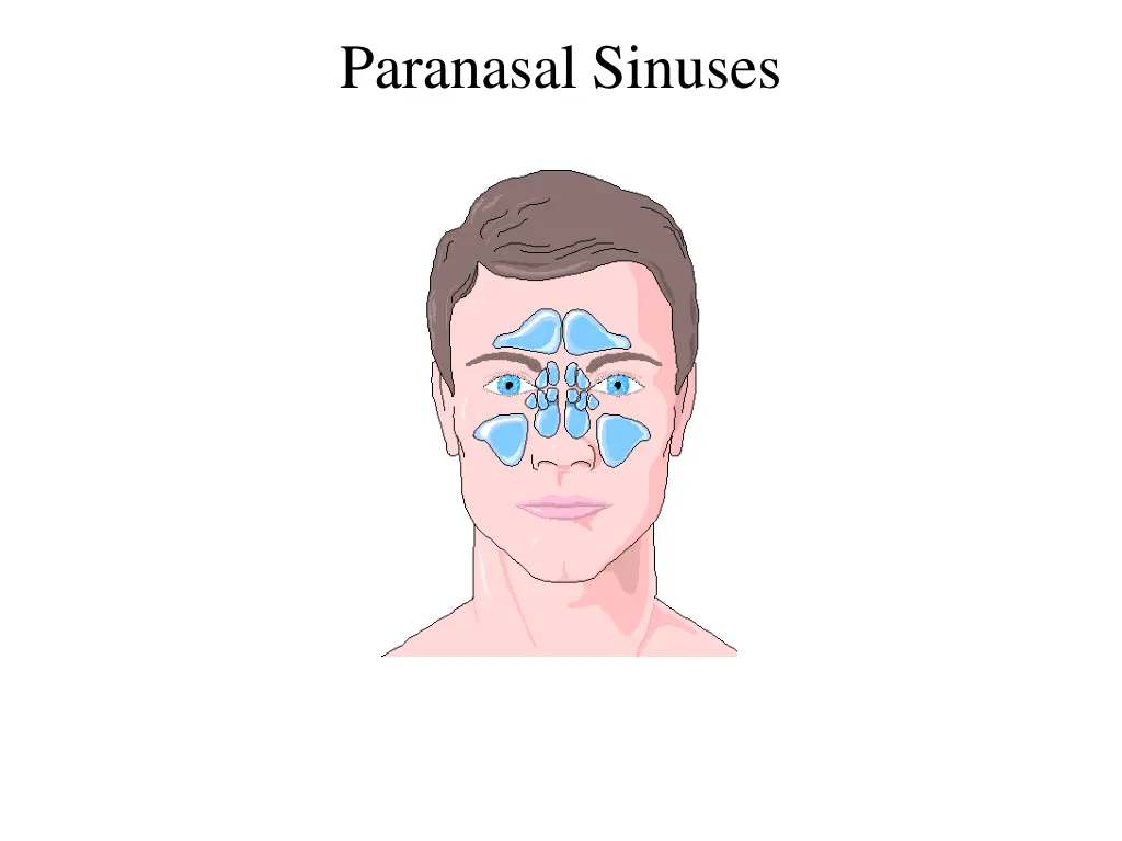
Introduction The paranasal sinuses are air-filled spaces located within the bonesof the skull and facial bones.The sinus is regarded by some as an accessory space to the nasal cavity, occurring only as a result of an inadequate process of ossification. In contrast, others report the functional contributions of the maxillary sinus in many aspects of olfactory and respiratory physiology. The sinuses are rudimentary or even absent at birth.They enlarge rapidly during the ages of six to seven years, i.e. time of eruption of permanent teeth and then after puberty. From birth to adult life the growth of the sinuses is due to enlargement of the bones; in old age it is due to resorption of the surrounding cancellous bone.
Types of paranasal sinuses - There are 4 pairs of paranasal sinuses 1) Frontal sinus 2) Maxillarysinus 3) Sphenoidal sinus 4) Ethmoidalsinus
1) Maxillary Sinus Definition- The maxillary sinus is the pneumatic space that is lodged inside the body of the maxilla and that communicates with the environment by way of the middle nasal meatus and the nasal vestibule. Anatomy of the maxillary sinus was first described by the British surgeon and anatomist Nathaniel Highmore in 1651. It is also known as the Antrum of Highmore .
Develomental aspect Maxillary sinus is the first of the paranasal sinus to develop. 4thweek intra-uterine life dorsal portion of 1stPharyngeal arch forms the Maxillary process, which extends forward and beneath the developing eye to give rise to the maxilla.
Horizontal shift of the palatal shelves & fusion with one another. Nasal septum separates the oral cavity from the two nasal chambers. Influences further expansion of the lateral nasal wall & 3 wall begin to fold. 3 conchae & meatuses arise
3 Meatus Superior & Inferior Meatus Middle Meatus Expands immediately into lateral nasal wall in an inferior direction occupying more of the further maxillary body Remain as shallow depressions along the lateral nasal wall for first half of I.U. life
Development of sinus starts at 12 weeks as an evagination of the mucous membrane in the lateral wall of the middle meatus of the nose when the nasalepithelium invades the maxillary mesenchyme.
Age changes of Maxillary Sinus Tubular at birth Ovoid in childhood Pyramidal in adulthood
0-3 years 3-4 years 5-9 years Increase in width with facialgrowth Size : 27mm X 18mm X 17mm At birth filled with deciduous tooth germs n d Position : 2 deciduousmolar & 1stpermanent molar Growth corresponds to permanent teeth eruption Size : 7mm X 4mm X 4mm V olume 6-8ml 20thmonth posterior development 3rdyear : 1/2 adult size
9-12 years 12-15 years Old age Resorption of ridge with continued sinus pneumatization which leaves a thin layer of corticle bone separating the sinus mucosa from oral mucosa Antralfloor same level with nasal floor Floor of sinus 5- 12mm below nasal floor Size : 32mm X 33mm X 25mm V olume : 15-20ml Floor : 1stmolar< 2ndmolar < 2nd premolar
Anatomy of Maxillary Sinus Largest of the paranasal sinus Pyramidal shaped cavity within the body of the Maxilla The base of the pyramid forming the lateral nasal wall and apex at the root of the zygoma.
Roof of the antrum Formed by floor of the orbit and is transversed by the infraorbital nerves.It is flat and slopes slightly anteriorly and laterally.
Floor of the sinus Curved rather than flat formed by alveolar process of the maxilla and lies about 1cm below the level of the floor of the nose. Closely related to root apices of the maxillary premolar and molar.
Anteriorwall Formed by the facial surface of the maxilla. Extends from pyriform aperture anteriorly to zygomaticomaxillary suture & inferior orbital rim superiorly to alveolar process inferiorly. Convexity towards sinus thinnest in canine fossa Importantstructures - Infraorbital foramen Anterior superior alveolar nerve (ASA) Canine Fossa Middle superior alveolar nerve (MSA).
Posteriorwall Made of zygomatic and greater wing of sphenoid bone. Athin plate of bone separate the antral cavity from the infratemporal fossa. Thick laterally, thin medially. Important structures - Posterior superior alveolar nerve Maxillaryartery Pterygopalatine ganglion Nerve of pterygoid canal
Medial wall Bounded by the nasal cavity The opening of the sinus is closer to the roof and thus at a higher level than the floor. Formed by lateral nasal wall Inferior - nasal conchae Posterior- palatine bone Superior -uncinate process of ethmoid,lacrimalbone Contains doublelayer of mucous membrane(parsmembranacea) Important structures- Sinusostium Hiatussemilunaris Ethmoidal bulla Uncinate process Infundibulum
Ostium Opening of the maxillary sinus is called osteum. Ostium of the maxillary sinus is situated high up in medial wall and opens into the middle meatus of the nose in the lower part of the hiatus semilunaris. Lies above the level of nasal floor. The ostium lies approximately 2/3rd making drainage of the sinus inherently difficult. up the medial wall of the sinus, Blockage of the ostium can easily occur when there is inflammation of the mucosal lining of the ostium. An accessory ostium is also present behind the main ostium in 30% cases.
Neurovascular supply Arterial supply : By facial artery branch of external carotid artery. By infra orbital & greater palatine arteries branch of maxillary artery which is branch of external carotid artery. Venous: Toanterior facial vein & pterygoid plexus. Infection from the maxillary sinus may spread to involve cavernous sinus via any of its draining veins as the pterygoid plexus communicates with the cavernous sinus by emissary vein.
Neurovascular supply Nerve supply : Anterior superior alveolar nerve Middle superior alveolar nerve Posterior superior alveolar nerve Lymphatic Drain : The lymphatic drains in to submandibular lymph nodes.
Microscopic Features The epithelial layer of maxillary sinus lining is thinner than that of nasal cavity. Lined by Ciliated pseudostratified columnar epithelium derived from olfactory epithelium of middle nasal meatus Most numerous cells -Columnar ciliated cells Additional cells- Basal cells,Columnar non-ciliated cells,Goblet cells
CiliatedCells The cilia is composed of typical 9+1 pairs of microtubules & provide mobile apparatus to the sinus epithelium which moves the debris, microorganisms and the mucous film lining the epithelial surface of the sinus into the nasal cavity through the ostium.
GobletCells It is mucous synthesizing and secreting cells. It resembles an inverted wine glass with a short stack like basal end containing the nucleus and a swollen apical end containing mucin. It is an apocrine gland, i.e it pours its secretion through rupture of its apical cell membrane that get regenerated. So it has all the criteria of the synthesizing and secreting cells.
2) Frontal Sinus Situated in between inner and outer table of frontal bone Usually asymmetrical Pyramidal in shape with apex upwards and base is formed by the floor Boundaries Anterior wall-outer table of frontal bone Posterior wall- inner table of frontal bone separates the sinus from cranial cavity Floor- formed by thin bone separating sinus from orbit Medial wall- forms the septum between two frontal sinuses
Averagemeasurements: Heightt.: 3.2cm Width: 2.4cm, Depth: 1.6cm Capacity- 5-10 ML NEUROV ASCULAR SUPPL Y: Blood supply - Supra orbital artery Venous return -Anastomotic veins in supra orbital notch, connecting supra orbital and superior ophthalmic veins. Lymphatic drainage Submandibular nodes. Nerve supply - Supra orbital nerve(ophthalmic nerve)
3) Sphenoidal Sinus There are two sphenoidal sinuses in the sphenoid bone divided unequally by a thin bony septum Relations- Laterally- cavernous sinus containing 3rd,4th,5th,6thcranial nerves, internal carotid artery, optic nerve Superiorly- pituitary gland, optic chiasma, olfactory bulb, frontal lobe Inferiorly-nasopharynx and vidian nerve Posteriorly- brainstem, basilar artery
NEUROVASCULAR SUPPLY: Blood supply: Posterior ethmoidal and internal carotid arteries. Venous drainage: Pterygoid venous plexuses and cavernous sinus. Nerve supply: Posterior ethmoidal nerve and orbital branches of pterygopalatine ganglion. Lymphatic drainage: Retropharyngeal nodes
4)Ethmoidal Sinus Thin walled air cavities in the lateral masses of ethmoid bone. Relations - Roof- anterior cranial fossa lateral to cribriform plate Lateral wall- orbit, optic nerve,nasolacrimal duct separated by thin bone called lamina papyracea Inferior- maxillary sinus Posteriorly- sphenoid sinus Medially- superior and middle turbinate
Divided into three groups anterior, middle, posterior 1)ANTERIOR ETHMOIDALSINUS : Has 1 to 11 air cells. NEUROVASCULAR SUPPLY:Anterior ethmoidal nerve and vessels. L YMPHA TICS : Submandibular nodes. Some of the important anterior group cells includes a) Agger nasi cells b) Ethmoidal bulla c) Supraorbital cells d) Frontoethmoidal cells e) Haller cells
2)MIDDLE ETHMOIDALSINUS : Has 1 to 7 air cells. NEUROVASCULAR SUPPLY:Anterior ethmoidal nerve and vessels and orbital branches of pterygopalatine ganglion. LYMPHATICS : Submandibular nodes. 3)POSTERIOR ETHMOIDALSINUS : Has 1 to 7 air cells. NEUROV ASCULAR SUPPL Y: Posterior ethmoidal nerve and vessels and orbital branches of pterygopalatine ganglion. LYMPHATICS : Retropharyngealnodes. Onodi cells one of the most important air cell of this group.


 Maxillary Sinus")


 Frontal Sinus")

 Sphenoidal Sinus")

Ethmoidal Sinus")

MIDDLE ETHMOIDALSINUS :")