Comprehensive Guide to Spirometry: Indications, Interpretation, and More
Spirometry is a physiological test used for diagnosing and monitoring pulmonary function. It is crucial in assessing symptoms, evaluating impairment, and screening individuals at risk for lung diseases. This test plays a vital role in occupational medicine, especially in evaluating cardiopulmonary fitness for job demands and monitoring exposure to harmful agents. Learn about the types of spirometers, lung volumes, hygiene practices, reference values, and interpretation methods. Understand the primary and secondary prevention aspects of spirometry in medical surveillance programs, periodic evaluations, and workers' compensation settings.
Download Presentation

Please find below an Image/Link to download the presentation.
The content on the website is provided AS IS for your information and personal use only. It may not be sold, licensed, or shared on other websites without obtaining consent from the author. If you encounter any issues during the download, it is possible that the publisher has removed the file from their server.
You are allowed to download the files provided on this website for personal or commercial use, subject to the condition that they are used lawfully. All files are the property of their respective owners.
The content on the website is provided AS IS for your information and personal use only. It may not be sold, licensed, or shared on other websites without obtaining consent from the author.
E N D
Presentation Transcript
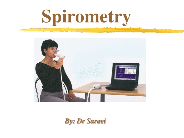
Spirometry By: Dr Saraei
Content Indication Indications in occupational medicine Contraindications Confounding factors Complications Type of spirometer Lung volumes & Lung capacities Spirometric values Hygiene & infection control Spirometry steps Reference values Interpretation
Definition of spirometry A physiological test for measuring volumes inhaled or exhaled by an individual as a function of time
Indication Not a screening test for general population Diagnostic Monitoring Impairment evaluation Public health
Indication (diagnostic) Evaluation of symptoms and signs Measuring the effect of dis. on pulmonary function Screening individuals at risk for pulmonary dis. Assess preoperative risk
Indication (monitoring) Assess therapeutic intervention Monitor people exposed to injurious agents
Indications in occupational medicine Primary prevention (Pre-employment) Physical demands of a job require a certain level of cardiopulmonary fitness, eg, heavy manual labor or firefighting Respirator use can impose a significant burden on the cardiopulmonary systems, eg, use of a self- contained breathing apparatus, or prolonged use of certain negative-pressure masks under conditions of heavy physical exertion and/or heat stress Research (Respiratory hazards)
Secondary prevention Medical surveillance programs & periodic evaluation OSHA : asbestos, cadmium, coke oven emissions, or cotton dust respirator-wearers exposed to benzene, formaldehyde methylene chloride Silicosis Spirometry detect large changes over a short time or smaller changes cumulated over a longer observation period, it is not sensitive to small, short-term changes Tertiary prevention Follow-up spirometry Workers compensation setting
Contraindications Active hemoptysis Pneumothorax Unstable Cardiovascular status (6 w) Cerebral/Thoracic/Abdominal aneurysm Recent eye surgery Acute disorder that may interfere with performance (e.g, vomiting) Thoracic or abdominal surgery( 3 w) Recent CVA or pulmonary emboli Respiratory distress
Confounding factors Common cold (3 days ago) Severe respiratory infection (3w) Smoking( 1hr) Heavy food (1hr) Bronchodilator use
Complications Chest pain Syncope, dizziness Increased ICP Paroxysmal coughing Nosocomial infection Bronchospasm
Spirometry standards ATS (American Thoracic Society) ERS (European Respiratory Society)
Lung volumes TV :The volume of air inhaled & exhaled at each breath during normal quiet breathing IRV: The maximum amount of air that can be inhaled after a normal inhalation ERV: The volume of air that can be forcefully expired following a normal quiet expiration RV: The volume of air remaining in the lungs after a forceful expiration
Lung capacities TLC: The total volume of the lungs VC:The maximum amount of air that can be exhaled after the fullest inspiration possible IC :The maximum of air that can be inhale after end tidal position FRC: The amount of air remaining in the lungs after a normal quiet expiration
Lung Volumes 4 Volumes IRV 4 Capacities Sum of 2 or more lung volumes IC VC TV TLC ERV FRC RV RV 15
Tidal Volume (TV) Volume of air inspired and expired during normal quiet breathing IRV IC VC TV TLC ERV FRC RV RV 16
Inspiratory Reserve Volume (IRV) The maximum amount of air that can be inhaled after a normal tidal volume inspiration IRV IC VC TV TLC ERV FRC RV RV 17
Expiratory Reserve Volume (ERV) Maximum amount of air that can be exhaled from the resting expiratory level IRV IC VC TV TLC ERV FRC RV RV 18
Functional Residual Capacity (FRC) Volume of air remaining in the lungs at the end of a TV expiration IRV The elastic force of the chest wall is exactly balanced by the elastic force of the lungs IC VC TV TLC ERV FRC = ERV + RV FRC RV RV 19
Total Lung Capacity (TLC) Volume of air in the lungs after a maximum inspiration IRV IC VC TV TLC TLC = IRV + TV + ERV + RV ERV FRC RV RV 20
Spirometric values FVC (forced vital capacity) FEV1 (forced expiratory volume in 1 s) FEV1/FVC FEF25-75 (maximum midexpiratory flow) PEF (peak expiratory flow) VT curve FV curve
Normal values depends on: Age Height Kyphoscoliosis arm span (H=arm span/1.06) Gender Race Caucasian ATS recommended scaling factor of 0.88 to the Caucasian predicted FEV1 and FVC for African- American, Chinese, and Japanese subjects
Hygiene & infection control Hand washing Gloves Disposable mouth piece & nose clip Disinfection or sterilization of reusable mouth piece Extra precautions for patient with known transmissible infection
Subject maneuvers FVC maneuver Closed circuit Open circuit Well-fitting false teeth yes or no Sitting or standing Nose clip Procedure 1. Inhale compete & rapid 2. Exhale: with minimal hesitation blast not just blow keep going
maneuver evaluation Start of test criteria - Extrapolation volume(EV < 5% of FVCor 150 ml) -Time-to-PEF < 0.120 s End of test criteria - the subject cannot or should not continue - exhalation at least 6s (in children <10 yrs: at least 3s) - volume-time curve show no change in volume (<0.025 lit) for at least 1s In obstruction or older subjects more than 6s exhalation (till 15s)
a b c
Acceptability Start of test criteria End of test criteria Cough especially during first second Valsalva maneuver (glottis closure) Leak from the mouth Obstruction of the mouthpiece Extra breath during the maneuver At the most eight tests should be performed
Acceptable spirogram Acceptable spirogram
rainbow G
Reproducibility At least three acceptable maneuvers Maximum difference between the largest and next largest FVC and FEV1 = 150ml or 5% (If FVC <1lit, this value is 100ml)
Reproducibility Reproducibility
Flow chart of criteria Perform FVC Acceptability criteria 3 acceptable maneuvers Repeatability criteria Largest FVC and largest FEV1 Maneuver with largest FVC + FEV1 for other indices
Reference values Knudson (male/ female) NHANES III (race difference) ACOEM recommends that the NHANES III equations be considered for general use in the occupational setting ERS ATS
LLN FEV1 and FVC = 80% FEV1/FVC = 70-75% FEF25-75 = 50-60%
A. Normal: both the FVC and the FEV1/VC ratio are normal. Knee
B.Obstructive : FEV1/FVC , FEV1 TLC & RV or NL
The severity of the abnormality is graded: - % Pred FEV1 > 100 = May be a physiological variant - % Pred FEV1 < 80 and > 70 = Mild - % Pred FEV1 < 70 and > 60 = Moderate - % Pred FEV1 < 60 and > 50 = Moderately severe - % Pred FEV1 < 50 and > 35 Severe - % Pred FEV1 < 34 = Very severe
C. Restrictive: FEV1/FVC or NL FVC & FEV1 , TLC & RV
The severity of the abnormality might be graded as follows: - % Pred FVC < LLN and > 70 = mild - % Pred FVC < 70 and > 60 = Moderate - % Pred FVC < 60 and > 50 = Moderately severe - % Pred FVC < 50 and > 34 = Severe - % Pred FVC < 34 = Very severe
D.Mixed pattern: FEV1,FVC, FEV1/FVC< LLN OR FEV1,FVC <LLN,FEV1/FVC:NL
VC VC VC RV RV RV Normal Obstructive Restrictive
Early small airway obx FV curve :upward concavity FVC, FEV1, FEV1/FVC :NL FEF 25-75 ??? ATS states that FEF25-75% should not be used to diagnose small airway disease or to assess respiratory impairment
Probably normal spirogram Only FEF 25-75 No small airway disease (ATS) If FEV1/FVC is borderline airway obx Only FEV1/FVC FEV1> 100% Normal FVC>100%

