Craniopharyngioma: Types, Epidemiology, Clinical Features, and Radiographic Characteristics
Craniopharyngioma is a benign brain tumor with two distinct types - adamantinomatous and papillary. It has a bimodal distribution in terms of incidence, presenting peaks in both children and young to middle-aged adults. Common symptoms include headaches, visual disturbances, hormonal imbalances, and behavioral changes. Pathologically, the tumor arises from Rathke's cleft, with different histological appearances for each subtype. Radiographically, craniopharyngiomas typically have a significant suprasellar component with the potential to distort surrounding structures. Understanding these key aspects of craniopharyngioma is crucial for diagnosis and management.

Download Presentation

Please find below an Image/Link to download the presentation.
The content on the website is provided AS IS for your information and personal use only. It may not be sold, licensed, or shared on other websites without obtaining consent from the author. If you encounter any issues during the download, it is possible that the publisher has removed the file from their server.
You are allowed to download the files provided on this website for personal or commercial use, subject to the condition that they are used lawfully. All files are the property of their respective owners.
The content on the website is provided AS IS for your information and personal use only. It may not be sold, licensed, or shared on other websites without obtaining consent from the author.
E N D
Presentation Transcript
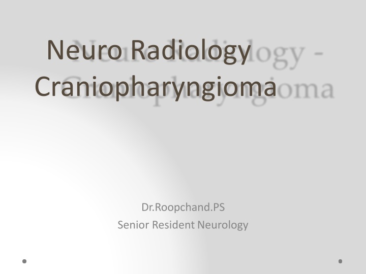
Neuro Radiology Craniopharyngioma Dr.Roopchand.PS Senior Resident Neurology
Introduction: Benign (WHO grade I) neoplasms which typically arise in the sellar / suprasellar region. Account for ~ 1 - 5% of primary brain tumours. Can occur anywhere from floor of the third ventricle, to the pituitary gland. Two pathological types and they differ in appearance, epidemiology and prognosis. o adamantinomatous (paediatric) o papillary (adult) o mixed: ~ 15%, but share imaging and prognosis similar to adamantinomatous
Epidemiology: Bimodal distribution: First peak between the ages of 10 - 14 years o Adamantinomatous type. Second peak in young to middle-aged adults o Papillary type Similar incidence in males and females.
Clinical presentation: Headaches and raised ICP Visual symptoms o 20% of children o 80% adults Hormonal imbalances o short stature and delayed puberty in children o decreased libido o amenorrhoea o diabetes insipidus Behavioural change due to frontal or temporal extension.
Pathology: Arises from the Rathke s cleft. This histological appearances of the two subtypes are different. Adamantinomatous: o In children o Reticular epithelial cells which have appearances reminiscent of the enamel pulp of developing teeth. o single or multiple cysts filled with thick oily fluid high in protein, blood products, and/or cholesterol, creating the so called "machinery oil". o "Wet keratin nodules" are a characteristic histological feature. o Calcification is usually present : ~ 90%
Papillary: o Seen almost exclusively in adults o Formed of masses of metaplastic squamous cells . o "Wet keratin" is absent. o Cysts do form, but these are less of a feature, and the tumour is more solid. o Calcification is uncommon or even rare
Radiographic features: Significant suprasellar component (95%), involving both the suprasellar and intrasellar spaces (75%). Purely suprasellar (20%), Purely intrasellar location is quite uncommon (<5%). Larger tumours can extend in all directions, frequently distorting the optic chiasm, or compressing the midbrain with resulting obstructive hydrocephalus. Occasionallycan appear as intraventricular, homogeneous, soft- tissue masses without calcification (papillary sub type). The third ventricle is a particularly common location. Rare / ectopic locations include: nasopharynx, posterior fossa, extension down the cervical spine.
Adamantinomatous: Lobulated contour as a result of usually multiple cystic lesions. Solid components are present. o Form a relatively minor component of the mass, Enhance vividly on both CT and MRI. Calcification is very common, but this is only true of the adamantinomatous subtype (90% are calcified) Predilection to be large, extending superiorly into the third ventricle, and encasing vessels, and even being adherent to adjacent structures.
CT cysts o typically large and a dominant feature o near CSF density solid component o soft tissue density o vivid enhancement calcification o seen in 90% o typically stippled and often peripheral in location
MRI cysts: variable but ~80% are mostly or partly T2 hyperintense solid component o T1: iso to lightly hypointense to brain o T1 C+: vivid enhancement o T2: variable / mixed calcification o difficult to appreciate on conventional imaging o susceptible sequences may better demonstrate calcification MR angiography: may demonstrate displacement of the A1 segment of the anterior cerebral artery MR spectroscopy: cyst contents may show a broad lipid spectrum, with an otherwise flat baseline 6
T1 T1C
Papillary : Papillary craniopharyngiomas tend to be more spherical in outline and usually lack the prominent cystic component. Most are either solid or contain a few smaller cysts. Calcification is uncommon or even rare in the papillary subtype
CT cysts o small and not a major feature o near CSF density solid component o soft tissue density o vivid enhancement calcification o uncommon - rare
MRI: cysts o when present they are variable in signal o 85% T1 hypointense solid component o T1: iso to lightly hypointense to brain o T1 C+: vivid enhancement o T2: variable / mixed MR spectroscopy: cyst contents does not show a broad lipid spectrum as they are filled with water fluid
Treatment is usually surgical with radiotherapy especially useful for incomplete resection. Benign local recurrence is seen in up to a third of patients. o papillary has a much lower recurrence rate than adamantinomatous Differentials o Rathke s cleft cyst. o Pituitary macroadenoma o Intracranial terratoma.