Critical Concepts in Respiratory Failure Management
This course delves into the definition of respiratory failure, common causes, clinical signs and investigations, differences in adults and children, clinical decision-making, and laboratory investigations including arterial blood gas interpretation. Learn key concepts to improve outcomes in pediatric respiratory failure.

Download Presentation

Please find below an Image/Link to download the presentation.
The content on the website is provided AS IS for your information and personal use only. It may not be sold, licensed, or shared on other websites without obtaining consent from the author.If you encounter any issues during the download, it is possible that the publisher has removed the file from their server.
You are allowed to download the files provided on this website for personal or commercial use, subject to the condition that they are used lawfully. All files are the property of their respective owners.
The content on the website is provided AS IS for your information and personal use only. It may not be sold, licensed, or shared on other websites without obtaining consent from the author.
E N D
Presentation Transcript
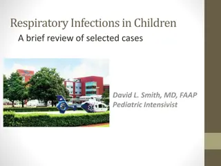
RESPIRATORY FAILURE RESPIRATORY FAILURE IN CHILDREN IN CHILDREN Critical Concepts Course
Objectives... Define respiratory failure Common causes of hypoxemia/hypercapnia Clinical signs/investigations
How is respiratory failure defined? Historically PaO2 <60 mm Hg, PaCO2 > 50 mm Hg Obviously must take into account patient s anatomy (ie - cyanotic heart lesion) Can develop acutely or over days How the patient looks is usually incorporated into diagnosis/management Symptoms/Severity dependent on acuity
Adults vs. Kids Multiple differences from underlying airway anatomy to disease process Kids usually affected by congenital or infectious processes Adults inflicted by respiratory disease such as COPD, as well as infectious processes Review differences in vital sign normals such as resp. rate, HR etc for children of different ages
Clinical decision making Acute vs. Chronic Helps in deciding acuity of treatment Progression of illness also important - history Any underlying chronic disease? i.e. Asthma, congenital heart disease Examine patient!!! Work of breathing, Level of consciousness, Vitals What tests might be helpful
Laboratory investigations Arterial blood gas (if possible) Gives info on oxygenation and ventilation status Difficult to get in some patients Obtaining and ABG should be part of resident skills Other blood gas ventilation info but not oxygenation Venous good only if obtained from free flowing site no tourniquet capillary easiest to obtain Other blood work based on clinical scenario (ie WBC count, cultures if suspect infection)
Important points on blood gas interpretation Know type of gas (ABG vs VBG vs CBG) Only interpret PaO2 on ABG PaCO2 slightly higher in VBG Remember metabolic side (base deficit, [HCO3-])
Oxyhemoglobin dissociation curve Two key points on curve: 1. PO2 100 mm Hg= SpO2 of 97% 2. PO2 40 mm Hg= SpO2 of 75% (mixed venous blood) Note the steep part of the curve in this area Small changes in clinical status will produce large swing in SpO2
Key points about the oxyhemoglobin saturation curve Remember how flat the slope is above PO2=60 mm Hg Any small drop in PO2 below this will cause precipitous fall in saturation
Oxygenation failure: Most common type of respiratory failure Occurs in wide variety of disease processes Main pathophysiologic derangements: V/Q mismatch Shunt Hypoventilation I. II. III.
Hypoventilation FiO2 of air is 21% PaO2 of air is (.21 X (760 mm Hg - 47 mm Hg (water vapor)) PO2 of alveolar gas is balance of removal and replenishment O2 consumption varies little Therefore, alveolar PO2 is determined mostly by level of alveolar ventilation If ventilation falls, PO2 drops and PCO2 will rise (this is key, hypoventilation will always lead to high PaCO2
Shunting Blood entering the arterial system without entering ventilated lung Intra- vs. extra-cardiac shunting Always a small amount of shunt via bronchial vessels, coronary veins Most important feature is 100% O2 does not resolve hypoxemia PCO2 usually normal or low as minute ventilation usually increased by chemoreceptors
Ventilation Perfusion Mismatch Ventilation / Blood flow are mismatched in different lung fields Most common cause of hypoxemia Usually exclude other causes before settling on V/Q mismatch
Ventilation Perfusion Mismatch Think of V/Q ratios varying from little to no ventilation (V/Q=0) to little to no blood flow (V/Q=infinity) Those lung units with low V/Q ratios cause hypoxemia Units with high V/Q ratios do not compensate for low O2 content of others due to shape of dissociation curve
Low V/Q unit with low end- capillary O2 content High V/Q unit with high end-capillary O2 content NOTE: Steep part of curve in range of low VO2 units PO2 = 70 mm Hg
VQ mismatch continues... Mismatch occurs in healthy lungs, difference is accounted for by regional blood flow/ventilation Ventilation / Perfusion both increase slowly from top to bottom of the lung Blood flow increases more rapidly than ventilation VQ ratio subsequently different as you move from 1 lung segment to the other Lungs with significant VQ mismatch cannot sustain the same levels of PaO2 /PaCO
What are the important clinical points? Is there an oxygenation defect? Check A-a gradient = PAO2 - PaO2(arterial) PAO2 = FiO2 - (PaCO2/0.8) (alveolar gas equ n) Normal value 5-30 mm Hg(age dependent) If elevated then almost always V/Q mismatch
Clinical examples of V/Q imbalance Asthma Pulmonary edema ARDS
How do you follow response to therapy? Options include: PaO2/FiO2 ratio Oxygenation index (OI) = Mean airway pressure (MAP) X FiO2 X 100% PaO2 Both validated but OI better when ventilated with positive pressure
Relationship between V/Q mismatch and gas exchange NOTE: Steep rate of decline in PaO2 compared to PaCO2
CO2 and respiratory failure Ventilation = the air moving in and out of lungs Minute ventilation is amount moving in and out per minute (VE) Alveolar ventilation is the volume of air that takes part in gas exchange. Dead space ventilation does not take part in ventilation PaCO2 is only measurement that reflects alveolar ventilation and the relationship to CO2 production CO2 production is continuous, elimination is through lungs predominantly
Why we care about hypoxemia/hypercarbia? Hypoxemia: Significant hypoxemia can lead to tissue hypoxia and anaerobic metabolism Different organ systems have different thresholds for tolerating hypoxemia (CNS and heart most vulnerable) Arterial PO2 is only one component of oxygen delivery (DO2), other important factors include hemoglobin level, cardiac output Rising serum lactate is an indicator of significant tissue hypoxia
Hypercarbia: Controversial topic with emergence of permissive hypercapnia in treatment of ALI/ARDS Definite CNS effects such as narcosis, mental clouding at high levels Adverse effects of acidosis produced by hypercarbia may be overstated Has demonstrated some protective effects against mechanical ventilation induced lung damage
In conclusion Think in terms of oxygenation and ventilation Think WHY (ie physiology) the patient is hypoxic/hypercarbic Remember to follow patients closely as they can deteriorate quickly
References, Recommended Reading, and Acknowledgements Up to Date: Emergent Evaluation of Acute Respiratory Distress in Children Nelson s Textbook of Pediatrics Some slides based on work by Dr. Jeff Brusinski for PedsCCM American Heart Association PALS guidelines