Cryptosporidium Parvum: Causes, Symptoms & Diagnosis
Learn about Cryptosporidium parvum, a microscopic parasite that causes cryptosporidiosis. Explore its classification, life cycle, sites of infection, and laboratory diagnosis methods. Understand the symptoms, especially in individuals with weakened immune systems, and discover how to identify the parasite in laboratory settings.

Download Presentation

Please find below an Image/Link to download the presentation.
The content on the website is provided AS IS for your information and personal use only. It may not be sold, licensed, or shared on other websites without obtaining consent from the author. If you encounter any issues during the download, it is possible that the publisher has removed the file from their server.
You are allowed to download the files provided on this website for personal or commercial use, subject to the condition that they are used lawfully. All files are the property of their respective owners.
The content on the website is provided AS IS for your information and personal use only. It may not be sold, licensed, or shared on other websites without obtaining consent from the author.
E N D
Presentation Transcript
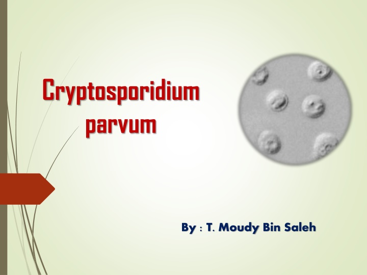
Cryptosporidium parvum By : T. Moudy Bin Saleh
Classification Phylum: Apicomplexa Class: Conoidasida Order: Eucoccidiorida Family: Cryptosporidiidae Genus: Cryptosporidium Species: C. parvum
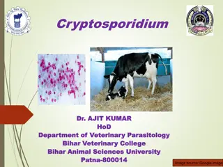
Causal Agents Cryptosporidiosis (or "Crypto" for short) is a disease that causes watery diarrhea. It is caused by microscopic germs parasites called Cryptosporidium. Although Crypto can affect all people, some groups are likely to develop more serious illness. For people with weakened immune systems, symptoms can be severe (life-threatening illness). For example: people with AIDS and cancer who are taking certain immunosuppressive drugs.
Site of infection Most parasite species infect the small intestines of their hosts (mammals) whereas others infect the respiratory tract (birds) or stomach (reptiles). The parasites are located within parasitophorous vacuoles covered by host microvillous membranes.
Life cycle They undergo cyclic asexual merogony (schizogony) followed by sexual gamogony ( microgametes fertilize macrogametes) resulting in the formation of small oocysts which undergo exogenous sporulation (forming 4 naked sporozoites). They undergo several cycles of asexualmerogonous development before gamonts are formed. After fertilization, the oocysts mature in the gut and are usually infective as soon as they are excreted from the host.
Laboratory Diagnosis Acid-fast staining methods, with or without stool concentration, are most frequently used in clinical laboratories. For greatest sensitivity and specificity, immunofluorescence microscopy is the method of choice (followed closely by enzyme immunoassays). Molecular methods are mainly a research tool.
Laboratory Identification of Parasitic Diseases Cryptosporidium parvum oocysts in wet mount, under differential interference contrast (DIC) microscopy. The oocysts are rounded and measure 4.2 m - 5.4 m in diameter. Sporozoites are visible inside the oocysts, indicating that sporulation has occurred. Cryptosporidium sp. oocysts stained with modified acid-fast
Thank you Thank you