Cyber Security Export and ITAR Brief 4 April 2018
In this briefing, delve into the challenges faced in higher education regarding cyber security, highlighting vulnerabilities, threats, and the importance of security awareness. Explore the risks associated with an open network, limited resources, and the need for compliance with regulatory standards like HIPAA, PII, PCI, GLBA, FISMA, FERPA, and ITAR. Discover the diverse range of security threats posed by nation states, terrorists, criminals, crackers, hackers, competitors, and script kiddies. Gain insights into the security vision for universities, addressing the protection of data, resources, and the digital transformation in cyber security.
Download Presentation

Please find below an Image/Link to download the presentation.
The content on the website is provided AS IS for your information and personal use only. It may not be sold, licensed, or shared on other websites without obtaining consent from the author.If you encounter any issues during the download, it is possible that the publisher has removed the file from their server.
You are allowed to download the files provided on this website for personal or commercial use, subject to the condition that they are used lawfully. All files are the property of their respective owners.
The content on the website is provided AS IS for your information and personal use only. It may not be sold, licensed, or shared on other websites without obtaining consent from the author.
E N D
Presentation Transcript
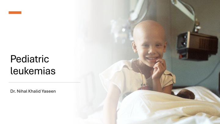
Pediatric leukemias Dr. Nihal Khalid Yaseen
To understand the incidence of leukemia in pediatrics. To recognize various types of leukemia in pediatrics with their clinical presentation and pathology. To outline a diagnostic approach to a patient with newly diagnosed leukemia. To recognize treatment outline of leukemia in infants and children with main treatment complications. To identify survivorship care plan. Learning objectives
Introduction Pediatric leukemia is a malignant diseases in which genetic abnormalities in a hematopoietic cell give rise to an unregulated clonal proliferation of cells. Leukemias are the most common malignant neoplasms in childhood, accounting for approximately 31% ( one third) of all malignancies in children younger than 15 years old. Nearly 80% of cases are ALL (acute lymphoblastic leukemia), 15% AML (acute myelogenous leukemia), 2% acute leukemia of ambiguous lineage, 2-3% CML (chronic myelogenous leukemia), and 1-2% JMML (juvenile myelomonocytic leukemia).
GENETIC SUSCEPTIBILITY Congenital syndromes: Down syndrome, Fanconi anemia, ataxia telangiectasia, NF-1, Kostmann syndrome Identical twins (higher risk of developing leukemia in a second member) Pesticide exposure, ionizing radiation, childhood infections, prior use of alkylating chemotherapeutic agents. 3-4 cases per 100,000 children in united states. Peak incidence between 2 and 5 years of age and more common in boys than in girls. The etiology of ALL is unknown, although several genetic and environmental factors are associated with childhood leukemia as shown in table (1). ENVIRONMENTAL FACTORS
FAB (French- American- British) classification according to cellular morphology where ALL is subclassified into L1, L2, L3 and AML is further subclassified into M0, M1, M2, M3, M4, M5, M6, & M7. Immunophenotypic classification according to surface markers on blast cells either: ALL which is subclassified into B- lymphoblastic leukemia (B cell origin) which constitutes 85% of cases and T- lymphoblastic leukemia which forms only 15% of cases, AML, or acute leukemia of ambiguous lineage. The World Health Organization (WHO) classification of ALL is based on cytogenetic and molecular characteristics of blast cells the most famous of these is the BCR ABL fusion gene formed by t(9;22) also termed as Philadilphia chromosome found in most cases of CML and certain cases of ALL also.
CLINICAL MANIFESTATIONS The initial presentation of ALL usually is nonspecific and relatively brief. Common symptoms are:- anorexia, fatigue, malaise, irritability, and fever are often present. Bone or joint pain, particularly in the lower extremities, may be present. Signs and symptoms of bone marrow failure as pallor, exercise intolerance, bruising, oral mucosal bleeding or epistaxis. Organ infiltration can cause lymphadenopathy, hepatosplenomegaly, testicular enlargement, or central nervous system (CNS) involvement (cranial neuropathies, headache, seizures, papilledema), ocular involvement may cause blurring of vision from leukemic infiltrates or retinal hemorrhages Respiratory distress may be caused by severe anemia or mediastinal node compression most frequently seen in adolescent males with T- cell ALL.
Patients with AML may present with:- subcutaneous nodules or blueberry muffin lesions (especially in infants), infiltration of the gingiva, signs and laboratory findings of disseminated intravascular coagulation (especially indicative of APL), and discrete masses, known as chloromas.
DIFFERENTIAL DIAGNOSIS Pancytopenia:- aplastic anemia (congenital or acquired), marrow infiltration from metastatic disease. Thrombocytopenia:- immune thrombocytopenia (ITP). Infections:- infectious mononucleosis (fever and lymphadenopathy), CMV infection. Rheumatological:- juvenile idiopathic arthritis (fever and bone pain)
Diagnosis Peripheral blood findings (CBC): anemia and thrombocytopenia are seen in most patients. Total WBC count may be low, normal or high, sometimes WBC count may be > 100*10 cells termed as hyperleukocytosis which causes symptoms from hyper viscosity and stasis of blood as CVA, bleeding, and respiratory acidosis. Blood film examination may show blast cells. Bone marrow examination: used to confirm the diagnosis. ALL is diagnosed by a bone marrow evaluation that demonstrates >25% of the bone marrow cells as a homogeneous population of lymphoblasts while the diagnosis of AML requires the presence of >20% myeloblasts in bone marrow aspiration or biopsy. Immunophenotyping using flow cytometry to identify subtype. Cytogenetics karyotype and molecular studies. CSF examination. If blasts are found and the CSF leukocyte count is elevated, overt CNS or meningeal leukemia is present.
Further workup Chest radiograph: Mediastinal mass more common in T-cell leukemia. Blood chemistry: Electrolytes, Blood urea nitrogen (BUN), creatinine, calcium, phosphorous, uric acid, Lactate dehydrogenase (LDH), and liver function tests. Patients may present with laboratory or clinical evidence of tumor lysis syndrome due to rapid lysis of malignant blasts leading to hyperuricemia, renal impairment, hypocalcemia, and hyperphosphatemia. Coagulation profile: Prothrombin time (PT), Partial Thromboplastin time (PTT), and fibrinogen.
Treatment The single most important prognostic factor in ALL is the treatment Risk-stratified therapy is the standard of current ALL treatment
Phases of ALL treatment Induction, aims to eradicate the leukemic cells from the bone marrow. Usually continues for 4 weeks and consists of vincristine, prednisone, and L-asparaginase. Patients at higher risk also receive daunorubicin. CNS prophylaxis using intrathecal methotrexate. Consolidation, focuses on intensive CNS therapy in combination with continued intensive systemic therapy to prevent later CNS relapses. Maintenance phase of therapy, which lasts for 2- 3 years. Patients are given mercaptopurine and methotrexate orally, usually with intermittent doses of vincristine. A small number of patients with particularly poor prognostic features, may undergo bone marrow transplantation during the first remission. Philadelphia chromosome positive ALL receive imatinib; an agent specifically designed to inhibit the BCR- ABL kinase resulting from the translocation.
For AML, various induction chemotherapy regimens exist, typically including an anthracycline (as Daunorubicin) in combination with high- dose cytarabine. Acute promyelocytic leukemia (APL), characterized by a gene rearrangement involving the retinoic acid receptor [t(15;17); PML- RARA], is very responsive to all- trans- retinoic acid (ATRA, tretinoin) combined with anthracyclines and cytarabine.
SUPPORTIVE CARE Patients with high WBC counts are especially prone to tumor lysis syndrome as therapy is initiated. The kidney failure associated with very high levels of serum uric acid can be prevented or treated with allopurinol or urate oxidase in addition to aggressive hydration using twice maintenance (3000ml/BSA). Chemotherapy often pro duces severe myelosuppression, which can require blood and platelet transfusion and aggressive empirical antimibiotics for sepsis in febrile children with neutropenia. Patients must receive prophylactic sulfamethoxazole for Pneumocystis jiroveci pneumonia during chemotherapy. Trimethoprim-
Improvements in therapy and risk stratification have resulted in significant increases in survival rates, with current data showing overall 5- year survival of ALL approximately 90%. The most important prognostic factors for ALL are shown in table 2. Patients with AML recently achieved survival rates of 60-70%. However, those with APL (acute promyelocytic leukemia) show excellent survival with the addition of ATRA to their treatment regimens. Also, patients with Down s syndrome have better prognosis due to greater sensitivity to chemotherapy. PROGNOSIS
Criteria Good prognosis Bad prognosis Age (yr) 1-10 <1 , >10 Gender Female Male Race White Black, Hispanics Immunophenotype B cell T cell CNS involvement No Yes Initial WBC count *10 /L <50 >50 Response to initial treatment as assessed by BMA Persistent minimal residual disease Low or absent minimal residual disease Favorable cytogenetics Present Absent
Survivorship care plan Survivors of childhood leukemia remain at risk for multiple long-term effects of therapy, including cardiac complications (Daunorubicin), neurocognitive issues (cranial irradiation), metabolic disorders, secondary (cyclophosphamide). They require close monitoring and follow-up. endocrine obesity, malignancies and and
References Nelson textbook f pediatric, 22nd edition, 2024 Lanzkowsky s manual of pediatric hematology and oncology, 7th edition, 2022 UpToDate
