Cysts of the jaws
A cyst in the jaws is a pathologic cavity filled with fluid, lined by epithelium, and surrounded by a definite CT wall. Radiographic features, internal structure, types of odontogenic and nonodontogenic cysts, and specifics on radicular cysts are discussed in detail.
Download Presentation

Please find below an Image/Link to download the presentation.
The content on the website is provided AS IS for your information and personal use only. It may not be sold, licensed, or shared on other websites without obtaining consent from the author.If you encounter any issues during the download, it is possible that the publisher has removed the file from their server.
You are allowed to download the files provided on this website for personal or commercial use, subject to the condition that they are used lawfully. All files are the property of their respective owners.
The content on the website is provided AS IS for your information and personal use only. It may not be sold, licensed, or shared on other websites without obtaining consent from the author.
E N D
Presentation Transcript
Definition A cyst is a pathologic cavity filled with fluid, lined by epithelium and surrrounded by a definite CT wall Cystic fluid either is secreted by the cells lining the cavity or derives from the surrounding tissue fluid
Radiographic features Location: centrally within maxilla and mandible and rare in coronoid and condylar process Found in tooth bearing region In mandible, originate above IAC Periphery: well defined and corticated In secondary infection or chronic state, changes to thicker, more sclerotic boundary Shape: round or oval, some have a scalloped boundary
Internal structure: totally rlucent Long standing cases may have dystrophic ossification Some may have septa Effects on surrounding structures: cysts grow slowly sometimes causing displacement and resorption of teeth Area of tooth resorption often has a sharp, curved shape Cysts can expand mandible, usually in smooth, curved manner May displace IANC in inferior direction or invaginate maxillary antrum, maintaining a thin layer of bone
Odontogenic cysts Radicular cyst Residual cyst Dentigerous cyst Buccal bifurcation cyst Odontogenic keratocyst Basal cell nevus syndrome Lateral periodontal cyst Calcifying odontogenic cyst
Nonodontogenic cyst Nasopalatine duct cyst Nasolabial cyst Dermoid cyst Cyst like lesions: Simple bone cyst
Radicular cyst Syn: periapical cyst, apical periodontal cyst, dental cyst 3rdto 6thdecade and slight male predominance Results when rests of ept cells (Malassez) in the periodontal ligament are stimulated to proliferate and undergo cystic degeneration by inflammatory products from a nonvital tooth C/F: are most common type of cyst in jaws Arise from nonvital tooth asymptomatic unless secondary infection occurs Swelling may feel bony hard if cortex is intact, crepitation as bone thins and rubbery and fluctant if cortex is lost
Radiographic features Epicentre: apex of a nonvital tooth Occasionally occurs on mesial or distal surface of root, at opening of accesory canal or infrequently in a deep periodontal pocket 60% are found in maxilla, splly around incisors and canines Periphery and shape: well defined corticated border If infection occurs, cortex is lost Outline is curved or circular
Occasionally dystrophic calcification may develop In long standing cases, and appear as sparsely distributed, small particulate r opacities Effects on surrounding structures: displacement and resorption of roots of adjacent teeth may occur Resorption pattern may have a curved pattern cyst may invaginate the antrum with evidence of a corticated boundary b/w contents of cyst and internal structure of antrum Outer paltes of maxilla and mandible may expand in a curved or circular shape IANC may dispalce in inferior direction Internal structure: radiolucent
Management Extraction, endo hterapy and apical surgery Larger cyst: surgical removal or marsupialization
Residual cyst Is a cyst that remains after incomplete removal of the original cyst C/F: asymptomatic cyst there may be some expansion of the jaw or pain in case of secondary infection r/g: occur in both jaws, more often seen in mandible Epicentre: periapical in location Periphery and shape: has a cortical margin unless it becomes secondary infected Shape is circular and oval
Internal structure: is radiolucent. Dystrophic calcifications may be seen in long standing cases Effects on surrounding structures: tooth dispalcement or resorption Outer cortical paltes of jaws may expand Cyst may invaginate the antrum or depress IANC Management: surgical removal or marsupialization
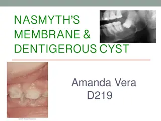
Dentigerous cyst Syn: follicular cyst A dentigerous cyst is a cyst that forms around the crown of an unerupted tooth It begins when fluid accumulates in layers of REE or b/w epithelium and crown of unerupted tooth Eruption cyst is soft tissue counterpart of dentigerous cyst C/F: are second most common type of cysts in jaws Develop around crown of unerupted or supernumerary tooth
Clinical examination reveals a missing tooth and possibly hard swelling, occasionally resulting in facial asymmetry Pt typically has no pain or discomfort R/G: Epicenter: just above the crown of involved tooth Cyst attaches at CEJ Some cysts are eccentric, developing from lateral aspect of follicle Periphery and shape: well defined cortex with a curved or circular outline If infection is present, cortex may be missing
Internal structure: completely rlucent Effects on surrouding structures: propensity to displace and resorb adjacent teeth It displaces associated tooth in an apical direction It may displace IANC in an inferior direction Expands the outer cortical boundary Floor of antrum may be dispalced as cyst invaginates antrum Management: surgical removal with tooth, large cyst with marsupialization
Buccal bifurcation cyst Syn: mandibular Infected buccal cyst, paradental cyst, inflammatory collateral dental cyst Source: epithelial cell rests in the periodontal membrane of the buccal bifurcation of mandibular molars C/F: common sign is the lack of or a delay in eruption of a mandibular first or second molar Molar may be missing or lingual cusp tips may be abnormally protruding through the mucosa, higher than position of buccal cusps
1stmolar is involved more frequently than 2ndmolar Teeth are always vital Hard swelling may be present buccal to involved molar Pt experiences pain if secondary infection is present Occurs within first 2 decades R/G: mandibular 1stmolar is most common location Followed by 2ndmolar It is occasionally bilateral Always located in buccal bifurcation of affected molar
Periphery and shape: lesion may be very subtle rlucent region superimposed over the image of roots of molar Lesion is circular in shape and well defined cortical border Internal structure: is r lucent Effects on surrounding structures: molar is tipped and so the root tips are pushed into lingual cortical plate of mandible Larger cyst may displace and resorb adjacent teeth and cause expansion
If cyst is secondarily infected, periosteal new bone formation is seen on buccal cortex adjacent to involved tooth Management: conservative curettage No recurrence
Odontogenic keratocyst Syn: primordial cyst Is a noninflammatory odontogenic cyst that arises from dental lamina C/F: wide age range Mostly during 2ndand 3rddecades Slight male predominance Cyst sometimes form around an unerupted tooth Asymptomatic, mild swelling may occur Pain may occur with secondary infection
r/g: location is posterior body of mandible and ramus Epicenter is above IANC Periphery and shape: cortical border, cyst may have a smooth round or oval shape or may have a scalloped border Internal structure: r lucent In some cases, curved internal seepta may be present Effects on surrounding structures: has the propensity to grow along the internal aspects of the jaws, causing minimal expansion
OKCs may displace and resorb teeth IANC may be displaced inferiorly In maxilla, it may invaginate and occupy the entire maxillary sinus Management: resection, curettage or marsupialization
Basal cell nevus syndrome Syn: nevoid basal cell carcinoma syndrome or Gorlin- Goltz syndrome It comprises a number of abnormalities such as multiple nevoid basal cell carcinomas of skin, skeletal abnormalities, CNS abnormalities, eye abnormalities and multiple OKC s C/F: starts to appear early in life usually after 5 yrs of age and before 30 yrs Lesion occur as multiple OKC s of jaws
Skin lesions are small, flattened, flesh-colored or brown papules that occur anywhere on body and are especially prominent on face, neck and trunk Skeletal abnormalities are bifid rib, and other coastal abnormalities such as agenesis, deformity and synostosis of ribs, kyphoscoliosis, vertebral fusion, polydactyly, shortening of metacarpals, temporal and temporoparietal bossing, minor hypertelorism and mild prognatism Calcification of the falx cerebri and other parts of dura occur early in life
Radiographic features Location: multiple OKC s may develop bilaterally and can vary in size from 1mm to several cms in diameter Radiopaque line of calcified falx cerebri may be prominent on PA skull projection Occasionally this calcification may occur laminated Management: treated more aggressively
Lateral periodontal cyst Are thought to arise from epithelial rests in periodontium lateral to tooth root C/F: lesions are usually asymptomatic and less than 1cm in diameter No sexual predilection 2ndto 9thdecade, mean age is 50 yrs R/G: occur mostly in the region of lateral incisor to 2ndpremolar in mandible Occasionally appear in maxilla, b/w lateral incisor and cuspid
Periphery and shape: well defined rlucecy with a prominent cortical boundary and a round or oval shape Rare large lesions have a more irregular shape Internal structure: radiolucent Botryoid variety have a multilocular appearance Effects on surrounding structures: small cysts may efface lamina dura of addjacent root Large cysts can displace adjacent teeth and cause expansion Management: excisional biopsy or simple enucleation
Calcifying odontogenic cyst Syn: calcifying epithelial odontogenic cyst or Gorlin cyst Are uncommon, slow growing benign lesions C/F: wide age range that peaks at 10-19 yrs of age, mean age of 36 yrs 2ndincidence peak in 7thdecade Lesion appears as a slow growing, painless swelling of jaw Occasionally pt c/o pain
In some cases, expanding laesion may destroy cortical plate and cystic mass may become palpable as it extends into soft tissue Pt may report a discharge from advanced lesions Aspiration yeilds a viscous, granular, yellow fluid R/G: equally distributed in both jaws Most occur anterior to 1stmolar, splly associated with cuspids and incisors
Periphery and shape: well defined and corticated with a curved, cyst like shape to illdefined and irregular Internal structure: completely r lucent It may show evidence of small foci of calcified material that appear as white flecks or small smooth pebbles or even larger, solid, amorphous massses Rarely lesion appear multilocular Effects on surrounding structures: cyst is associated with a tooth and impedes its eruption
Displacement of teeth and resorption of roots may occur Perforation of cortical plate may be seen with enlarging lesions Management: enucleation and curettage
Nasopalatine duct cyst Syn: nasoplatine canal cyst, incisive canal cyst, nasoplatine cyst, medial palatine cyst, median anterior maxillary cyst C/F: 4ththrough 6thdecades M:F::3:1 Asymptomatic or cause minor symptoms Frequent complaint is a small well defined swelling just posterior to palatine papilla Swelling is usually fluctuant and blue if cyst is near the surface
Deeper lesions have normal appearing mucosa If cyst expands, it may penetrate labial plate and produce a swelling below maxillary labial frenum Lesion also may bulge into nasal cavity and distort nasal septum Pressure from cyst on adjacent nerves may cause a burning sensation or numbness over palatal mucosa In some cases, cystic fluid may drain into oral cavity through a sinus tract or a remnant of nasopalatine duct Pt usually detects fluid and reports a salty taste
Radiographic features Location: nasopalatine canal or foramen If it extends posteriorly to involve hard palate, It is referred to as median palatal cyst If it extends anteriorly b/w central incisors, destroying or expanding labial plate of bone and causing the teeth to deverge, it is median anterior maxillary cyst Periphery and shape: well defined and corticated and is circular or oval Shadow of nasal spine is superimposed on cyst, giving it a heart shape
Internal structure: totally rlucent Rarrely, they amy have dystrophic calcifications, appearing as ill defined, amorphous, scattered r opacities Effects on surrounding structures: cyst causes the roots of central incisors to diverge Occasionally root resorption occurs
Cyst may expand the labial and palatal cortex Floor of nasal fossa may be displaced in a superior direction Management: enucleation
Nasolabial cyst Syn: nasoalveolar cyst Arise from epithelial rests in fusion lines of globular, lateral nasal and maxillary processes C/F: small lesion produce a very subtle, unilateral swelling of nasolabial fold and may elicit pain or discomfort If large, it may bulge into floor of nasal cavity, causing some obstruction, flaring of the alae,distortion of nostrils and fullness of upper lip If infected, may drain into nasal cavity Usually unilateral but bilateral lesions have occured
Mean age is 44 yrs 75% of lesions occur in females R/G: are primarily soft tissue lesions located adjacent to alveolar process above apices of incisors Periphery and shape: thin axial CT images using soft tissue algorithm with contrast reveal a circular or oval lesion with a slight soft tissue enhancement of periphery
Internal structure: homogenous and relatively r lucent compared with surrounding soft tissues Effects on surrounding structures: occasionally causes erosion of underlying bone, producing an increased r lucency of alveolar process beneath the cyst and apical to incisors The usual outline of inferior border of nasal fossa may become distorted, resulting in a posterior bowing of this margin Management: excised through intraoral approach
Dermoid cyst Are a cystic form of a teratoma thought to be derived from trapped embroynic cells that are totipotential C/F: may develop in the soft tissues at any time from birth Become apparent from 12 to 25 yrs of age Equally distributed b/w both sexes Swelling, which is slow and painless, can grow to several cms in diameter If located in tongue or neck, may interfere with breathing, speaking and eating Depending on the depth, it may deform the submental area
