Cytotoxicity and Adipocyte Response to Protease Inhibitors
Explore the cytotoxicity and adipokine secretion effects of protease inhibitors on 3T3-F442A and primary human adipocytes, showcasing lipid accumulation and gene expression changes. This study investigates the impact of telmisartan and rosiglitazone co-incubation in the adipocyte models. Discover the influence on cell viability, lipid accumulation, gene expression of PPAR and Lipin1, and adipokine secretion levels.

Download Presentation

Please find below an Image/Link to download the presentation.
The content on the website is provided AS IS for your information and personal use only. It may not be sold, licensed, or shared on other websites without obtaining consent from the author. If you encounter any issues during the download, it is possible that the publisher has removed the file from their server.
You are allowed to download the files provided on this website for personal or commercial use, subject to the condition that they are used lawfully. All files are the property of their respective owners.
The content on the website is provided AS IS for your information and personal use only. It may not be sold, licensed, or shared on other websites without obtaining consent from the author.
E N D
Presentation Transcript
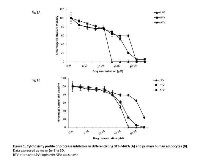
120 Fig 1A LPV Percentage Control cell Viability RTV 100 ATV 80 60 40 20 0 Drug concentration ( M) 120 Fig 1B LPV Percentage Control cell Viability RTV 100 ATV 80 60 40 20 0 Drug concentration ( M) Figure 1. Cytotoxicity profile of protease inhibitors in differentiating 3T3-F442A (A) and primary human adipocytes (B). Data expressed as mean (n=3) SD. RTV: ritonavir; LPV: lopinavir; ATV: atazanavir.
0.3 Absorbance of lipid bound Oil Red O Fig 2A 0.25 0.2 * * 0.15 (units) 0.1 0.05 0 0.6 Absorbance of lipid bound Oil Red O Fig 2B 0.5 0.4 (units) 0.3 * * 0.2 0.1 0 Figure 2. Lipid accumulation in differentiating 3T3-F442A (A) and primary human adipocytes (B) following incubation with protease inhibitors with/without telmisartan or rosiglitazone. Rosiglitazone was coincubated with lopinavir in the primary human adipocyte model only. *=P<0.01; Drug v Vehicle. =P<0.01; Drug v Drug+TEL or Drug+ROSI. Data expressed as mean (n=3) SD. Veh: vehicle; RTV: ritonavir; LPV: lopinavir; ATV: atazanavir; TEL: Telmisartan; ROSI: Rosiglitazone.
Fig 3A Fig 3C 1.2 ppar- relative gene expression 1.2 lpin1 relative gene expression 1 1 0.8 0.8 ( Ct) 0.6 ( Ct) 0.6 0.4 * * 0.4 * * 0.2 0.2 0 0 Fig 3B Fig 3D 1.2 1.2 PPAR- relative gene expression LPIN1 relative gene expression ( Ct) 1 1 0.8 0.8 ( Ct) 0.6 0.6 * 0.4 * 0.4 * * 0.2 0.2 0 0 Figure 3. Expression of PPAR (3A, 3T3-F442A; 3B, primary human adipocytes) and Lipin1 (3C, 3T3-F442A; 3D, primary human adipocytes) following incubation with protease inhibitors with/without telmisartan or rosiglitazone. Rosiglitazone was coincubated with lopinavir in the primary human adipocyte model only. Data expressed as mean (n=3) SD. *=P<0.01; Drug v Vehicle. =P<0.01; Drug v Drug+TEL or Drug+ROSI. =P<0.01; Preadipocyte v Vehicle. Preadipo: preadipocyte; Veh: vehicle; RTV: ritonavir; LPV: lopinavir; ATV: atazanavir; TEL: Telmisartan; ROSI: Rosiglitazone.
Fig 4A Fig 4C 20 Adiponectin protein (ng/ml) 300 18 * 16 IL-6 Protein (ng/ml) 250 14 * 200 12 10 150 * 8 100 6 * 4 50 2 0 0 Fig 4B Fig 4D 30 Adiponectin Protein (ng/ml) 300 * 25 * IL-6 Protein (ng/ml) 250 20 200 15 150 10 100 * 5 50 * 0 0 Figure 4 legend in the next slide
Fig 4E Fig 4G 70 * 0.8 60 TNF Protein (pg/ml) Resistin Protein (ng/ml) 0.7 * 50 0.6 40 0.5 0.4 30 * 0.3 20 * * 0.2 10 0.1 0 0 Fig 4F Fig 4H 90 * 80 0.9 TNF Protein (pg/ml) * 70 Resistin Protein (ng/ml) 0.8 60 0.7 0.6 50 0.5 40 * 0.4 30 * * 0.3 20 0.2 10 0.1 0 0 Figure 4. Adipokine secretion in differentiating 3T3-F442A and primary human adipocytes following incubation with protease inhibitors with or without telmisartan or rosiglitazone (Adiponectin: 4A, 3T3-F442A; 4B, primary human adipocytes; IL-6: 4C, 3T3-F442A; 4D, primary human adipocytes; TNF- : 4E, 3T3-F442A; 4F, primary human adipocytes; Resistin: 4G, 3T3-F442A; 4H, primary human adipocytes) Rosiglitazone was coincubated with lopinavir in the primary human adipocyte model only. Data expressed as mean (n=3) SD. *=P<0.01; Drug v Vehicle. =P<0.01; Drug v Drug+TEL or Drug+ROSI. =P<0.01; Preadipocyte v Vehicle. Preadipo: preadipocyte; Veh: Vehicle; RTV: ritonavir; LPV: lopinavir; ATV: atazanavir; TEL: Telmisartan; ROSI: Rosiglitazone.
120 Free fatty Acid released ( g/ml) Fig 5A * 100 * 80 60 40 20 0 Fig 5B 1.2 1 pAkt : Total Akt (Units) 0.8 0.6 * * 0.4 0.2 0 Figure 5. Lipolysis (A) and expression of pAktSer473 (B) in human primary adipocytes following incubation with protease inhibitors with or without telmisartan or rosiglitazone. Data expressed as mean (n=3) SD. pAkt expression was adjusted to Total Akt and data is expressed as mean ratio of absorbance. *=P<0.01; Drug v Vehicle. =P<0.01. Drug v Drug+TEL or Drug+ROSI. Veh: Vehicle; RTV: ritonavir; LPV: lopinavir; ATV: atazanavir; TEL: Telmisartan; ROSI: Rosiglitazone.
1.2 PPAR relative gene expression ( Ct) Fig 6A 1 0.8 0.6 0.4 * 0.2 0 45 Fig 6B 40 Adiponectin Protein (ng/ml) 35 30 25 20 15 10 * 5 0 Figure 6. Dose-response relationship between telmisartan and in vitro metabolic effects: Effect of telmisartan on PPAR (A) and secreted adiponectin (B) over full concentration range. Data expressed as mean (n=3) SD. *=P<0.01; Drug v Vehicle. =P<0.01; Drug v Drug+TEL or Drug+ROSI. Veh: Vehicle; LPV: lopinavir; TEL: Telmisartan; ROSI=Rosiglitazone.