Learn about dental calculus, its composition, formation, and etiologic significance, along with predisposing factors and the primary cause of gingival inflammation being bacterial plaque. Explore the types and characteristics of calculus and its impact on oral health.
Please find below an Image/Link to download the presentation.
The content on the website is provided AS IS for your information and personal use only. It may not be sold, licensed, or shared on other websites without obtaining consent from the author. If you encounter any issues during the download, it is possible that the publisher has removed the file from their server.
You are allowed to download the files provided on this website for personal or commercial use, subject to the condition that they are used lawfully. All files are the property of their respective owners.
The content on the website is provided AS IS for your information and personal use only. It may not be sold, licensed, or shared on other websites without obtaining consent from the author.
E N D
Presentation Transcript
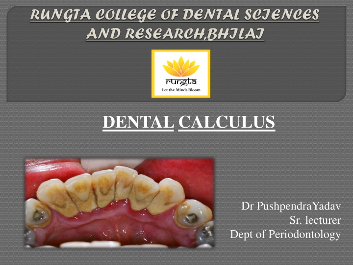
DENTALCALCULUS Dr PushpendraYadav Sr. lecturer Dept of Periodontology
CORE AREAS DOMAIN CATEGORY INTRODUCTION Affective Desire to know DEFINITION Cognitive Must to know CLASSIFICATION Cognitive Must to know COMPOSITION Cognitive Must to know FORMATION Cognitive Must to know THEORIES Cognitive Must to know ETIOLOGIC SIGNIFICANCE Cognitive Must to know MATERIA ALBA, FOOD DEBRIS, AND DENTAL STAINS Other predisposing factors Cognitive Must to know Cognitive Must to know FOOD IMPACTION Cognitive Must to know MATERIALS Cognitive Must to know PARAFUNCTIONAL HABIT Cognitive Must to know
PART I INTRODUCTION DEFINITION CLASSIFICATION COMPOSITION FORMATION PART II THEORIES ETIOLOGIC SIGNIFICANCE MATERIA ALBA, FOOD DEBRIS, AND DENTAL STAINS OTHER PREDISPOSING FACTORS FOOD IMPACTION MATERIALS PARAFUNCTIONAL HABIT SUMMARY REFERENCE
other predisposing factors include calculus, malocclusion, faulty restorations, complications associated with orthodontic therapy, self inflicted injuries, use of tobacco, and radiation therapy.
According to location a) Supragingival calculus b) Subgingival calculus According to source of mineralization a) Salivary calculus b) Serumal calculus According to surface a) Exogeneous b) Endogeneous
LOCATION: coronal to the gingival margin and therefore is visible in the oral cavity. COLOR: It is usually white or whitish yellow in color, CONSISTENCY: hard with claylike consistency, and easily detached from the tooth surface.
The color is influenced by contact with such substances as tobacco and food pigments. It may localize on a single tooth or group of teeth, or it may be generalized throughout the mouth.
The two most common locations: the buccal surfaces of the maxillary molars and the lingual surfaces of the mandibular anterior teeth
Saliva from the parotid gland flows over the facial surfaces of upper molars through Stensen s duct, whereas the orifices of Wharton s duct and Bartholin s duct empty onto the lingual surfaces of the lower incisors from the submaxillary and sublingual glands, respectively.
Location: below the crest of the marginal gingiva and therefore is not visible on routine clinical examination. evaluated by careful tactile perception with a an explorer. hard and dense and frequently appears dark brown or greenish black while being firmly attached to the tooth surface
When the gingival tissues recede, subgingival calculus becomes exposed and is therefore reclassified as supragingival . Thus, supragingival calculus can be composed of both supragingival calculus and previous subgingival calculus.
Both supragingival calculus and subgingival calculus may be seen on radiographs . Highly calcified interproximal calculus deposits are readily detectable as radiopaque projections that protrude into the interdental space
SUPRAGINGIVAL SUPRAGINGIVAL CALCULUS CALCULUS 1. It is located coronal to gingival margin 2. It is visible in the oral cavity SUBGINGIVAL SUBGINGIVAL CALCULUS CALCULUS Located below the crest of marginal gingiva. Not visible on routine clinical examination. May be found by tactile exploration, radiographs, jet air blowing . 3. Color - White or whitish yellow Dark brown or greenish black 4. Consistency - hard clay like Hard or flint like 5.Easily detached from tooth surface. 6.Also called salivary calculus Dense and is firmly attached to tooth surface Serumal calculus 7.Main mineral source - saliva. Main mineral source Gingival crevicular fluid.
Organic salivary glycoprotein 5.9% to 8.2% Carbohydrate 1.9% to 9.1% Lipids 0.2% galactose, glucose, rhamnose, mannose, glucuronic acid, galactosamine neutral fats, free fatty acids, cholesterol, cholesterol esters, and phospholipids sometimes arabinose, galacturonic acid, and glucosamine
Higher calcium :phosphate ratio Sodium content increases with the depth of periodontal pockets. Salivary proteins not found subgingivally
MORE Magnesium whitlockite SAME Hydroxyapatit e LESS LESS Brushite Octacalcium phosphate
Attachment by means of an organic pellicle on enamel Mechanical locking into surface irregularities, such as resorption lacunae Close adaptation of calculus undersurface depressions to the gently sloping mounds of the unaltered cementum surface Penetration of calculus bacteria into cementum calculocementum
between 1st and 14th days of plaque formation. Soft plaque is hardened by mineralization occur as soon as 4-8 hrs. Calcification Mineralization: 50% in 2 days and 60% to 90% in 12 days
The average daily increment in calculus formers is from 0.10% to 0.15% of dry weight. Calculus formation continues until it reaches a maximum, after which it may be reduced in amount. The time required to reach the maximal level has been reported as 10 weeks and 6 months.
The decline from maximal calculus accumulation, referred to as reversal phenomenon, may be explained by the vulnerability of bulky calculus to mechanical wear from food and from the cheeks, lips, and tongue.
Calculus can also occur readily in germ free animals. Calculocementum is the calculus embedded deeply in the cementum and which appears morphologically similar to cementum. Calculus is the most prominent plaque retentive factor and is a secondary etiologic factor for periodontitis.
Newman MG, Takei HH, Klokkevold PR, Carranza FA. Carranza s clinical periodontology, 10th ed. Saunders Elsevier; 2007. Lindhe J, Lang NP and Karring T. Clinical Periodontology and Implant Dentistry. 6th ed. Oxford (UK): Blackwell Publishing Ltd.; 2015. Newman MG, Takei HH, Klokkevold PR, Carranza FA. Carranza s clinical periodontology, 13th ed. Saunders Elsevier; 2018.