Explore the formation and composition of dental calculus, including factors contributing to its formation such as mineral precipitation and colloidal proteins. Learn about the impact of pH changes in saliva and the role of enzymes in calcium phosphate precipitation.
Please find below an Image/Link to download the presentation.
The content on the website is provided AS IS for your information and personal use only. It may not be sold, licensed, or shared on other websites without obtaining consent from the author. If you encounter any issues during the download, it is possible that the publisher has removed the file from their server.
You are allowed to download the files provided on this website for personal or commercial use, subject to the condition that they are used lawfully. All files are the property of their respective owners.
The content on the website is provided AS IS for your information and personal use only. It may not be sold, licensed, or shared on other websites without obtaining consent from the author.
E N D
Presentation Transcript
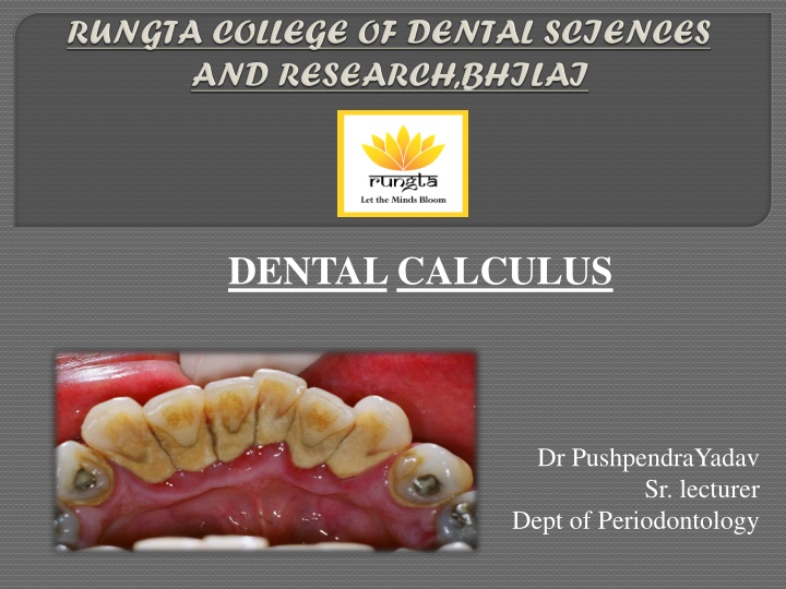
DENTALCALCULUS Dr PushpendraYadav Sr. lecturer Dept of Periodontology
CORE AREAS DOMAIN CATEGORY INTRODUCTION Affective Desire to know DEFINITION Cognitive Must to know CLASSIFICATION Cognitive Must to know COMPOSITION Cognitive Must to know FORMATION Cognitive Must to know THEORIES Cognitive Must to know ETIOLOGIC SIGNIFICANCE Cognitive Must to know MATERIA ALBA, FOOD DEBRIS, AND DENTAL STAINS Other predisposing factors Cognitive Must to know Cognitive Must to know FOOD IMPACTION Cognitive Must to know MATERIALS Cognitive Must to know PARAFUNCTIONAL HABIT Cognitive Must to know
PART I INTRODUCTION DEFINITION CLASSIFICATION COMPOSITION FORMATION PART II THEORIES ETIOLOGIC SIGNIFICANCE MATERIA ALBA, FOOD DEBRIS, AND DENTAL STAINS OTHER PREDISPOSING FACTORS FOOD IMPACTION MATERIALS PARAFUNCTIONAL HABIT SUMMARY REFERENCE
An increase in the pH of the saliva I causes precipitation of calcium phosphate salts The pH may be elevated by the... loss of carbon dioxide and the formation of ammonia by dental plaque bacteria or by protein degradation during stagnation.
Colloidal proteins in saliva bind calcium and phosphate ions II maintain a supersaturated solution with respect to calcium phosphate salts. With stagnation of saliva, colloids settle out, and the supersaturated state is no longer maintained, leading to precipitation of calcium phosphate salts
III Phosphatase liberated from dental plaque, desquamated epithelial cells, or bacteria precipitates calcium phosphate by hydrolyzing organic phosphates in saliva, thus increasing the concentration of free phosphate ions.
Esterase may initiate calcification by hydrolyzing fatty esters into free fatty acids. The fatty acids form soaps with calcium and magnesium later converted into the less soluble calcium phosphate salts.
The carbohydrateprotein complexes may initiate calcification by removing calcium from the saliva (chelation) and binding with it form nuclei induce subsequent deposition of minerals.
Filamentous organisms, diphtheroids, and Bacterionema and Veillonella species have the ability to form intracellular apatite crystals. Mineralization spreads until the matrix and the bacteria are calcified.
Calculus is always covered with a nonmineralized layer of plaque. A positive correlation between the presence of calculus and the prevalence of gingivitis exists, but this correlation is not as great as that between plaque and gingivitis. The initiation of periodontal disease in young people is closely related to plaque accumulation, whereas calculus accumulation is more prevalent in chronic periodontitis found in older adults.
The incidence of calculus, gingivitis, and periodontal disease increases with age. Calculus does not contribute directly to gingival inflammation, but it provides a fixed nidus for the continued accumulation of bacterial plaque and its retention in close proximity to the gingiva.
Plaque initiates gingival inflammation, which leads to pocket formation, and the pocket in turn provides a sheltered area for plaque and bacterial accumulation. The increased flow of gingival crevicular fluid associated with gingival inflammation provides the minerals that mineralize the continually accumulating plaque, resulting in the formation of subgingival calculus
The removal of subgingival plaque and calculus constitutes the cornerstone of periodontal therapy. Calculus plays an important role in maintaining and accentuating periodontal disease by keeping plaque in close contact with the gingival tissue and by creating areas where plaque removal is impossible.
Materia alba is an accumulation of microorganisms, desquamated epithelial cells, leukocytes, and a mixture of salivary proteins and lipids, with few or no food particles; it lacks the regular internal pattern observed in plaque. It is a yellow or grayish-white, soft, sticky deposit, and it is somewhat less adherent than dental plaque. The irritating effect of materia alba on the gingiva is caused by bacteria and their products.
Food Debris Most food debris is rapidly liquefied by bacterial enzymes and cleared from the oral cavity by salivary flow and the mechanical action of the tongue, cheeks, and lips. The rate of clearance from the oral cavity varies with the type of food and the individual. Aqueous solutions are typically cleared within 15 minutes, whereas sticky foods may adhere for more than 1 hour. Dental plaque is not a derivative of food debris, and food debris is not an important cause of gingivitis.
Dental stains Pigmented deposits on the tooth surface are called dental stains. primarily an esthetic problem and do not cause inflammation of the gingiva. The use of tobacco products, coffee, tea, certain mouthrinses, and pigments in foods can contribute to stain formation.
Iatrogenic factors Margins of restoration Contours and open contacts Materials Design of removable partial dentures Restorative dentistry procedures Malocclusion Parafunctional habits
Inadequate dental procedures that contribute to the deterioration of the periodontal tissues are referred to as iatrogenic factors. Eg: root perforations, vertical root fractures, and endodontic failures that may necessitate tooth extraction.
MARGINS OF RESTORATIONS Overhanging margins of dental restorations contribute to the development of periodontal disease by (1) changing the ecologic balance of the gingival sulcus to an area that favors the growth of disease-associated organisms (predominantly gram-negative anaerobic species) at the expense of the health-associated organisms (predominantly gram-positive facultative species) and (2) inhibiting the patient s access to remove accumulated plaque.
increase plaque accumulation, gingival inflammation, and the rate of gingival crevicular fluid flow. Subgingival at the level of the gingival crest induce less severe inflammation associated with degree of periodontal health similar to that seen with nonrestored interproximal surfaces. Supragingival
Roughness in the subgingival area is considered to be a major contributing factor to plaque buildup and subsequent gingival inflammation. The subgingival zone is composed of the margin of the restoration, the luting material, and the prepared and unprepared tooth surfaces.
Sources of marginal roughness include grooves and scratches in the surface of acrylic resin, porcelain, or gold restorations; separation of the restoration margin and the luting material from the cervical finish line, thereby exposing the rough surface of the prepared tooth; dissolution and disintegration of the luting material between the preparation and the restoration, thereby leaving a space; inadequate marginal fit of the restoration.
Overcontoured crowns and restorations tend to accumulate plaque and handicap oral hygiene measures in addition to possibly preventing the self-cleaning mechanisms of the adjacent cheek, lips, and tongue. Undercontoured crowns do not retain as much plaque as overcontoured crowns. Restorations that fail to reestablish adequate interproximal embrasure spaces are associated with papillary inflammation.
Definition: forceful wedging of food into the periodontium by occlusal forces. As the teeth wear down, their originally convex proximal surfaces become flattened, and the wedging effect of the opposing cusp is exaggerated.
Cusps that tend to forcibly wedge food into interproximal embrasures are known as PLUNGER CUSPS. These plunger cusp are usually the functional cusp (i.e. palatal cusp of maxillary teeth and buccal cusp of mandibular teeth) and sometimes palatal incline of maxillary buccal cusp & buccal incline of lingual cusp.
The forceful wedging of food normally is prevented by the: Integrity and location of proximal contact, The contour of the marginal ridge and developmental grooves, and The contour of the facial and the lingual surfaces.
According to Hirschfield (1930) Factors Causing Food Impaction Class I: Occlusal wear Class II: Loss of proximal contact Class III: Extrusion beyond the occlusal plane Class IV: Congenital morphological abnormality Class V: Improperly constructed restorations
1. DISCOMFORT A. Feeling of pressure B. Vague pain C. Root caries 2. PERIODONTAL CHANGES A. Gingival inflammation-bleeding & foul taste B. Gingival recession C. Periodontitis D. Periodontal abscess formation E. Alveolar bone loss - vertical
The use of rubber dam clamps, matrix bands, and burs in such a manner as to lacerate the gingiva results in varying degrees of mechanical trauma and inflammation. forceful packing of a gingival retraction cord into the sulcus
Bruxism..grinding of the teeth, excessive tooth wear Clenching when a person holds the teeth firmly together with significant force associated with masticatory system disorders
Favors plaque retention, by directly injuring the gingiva as a result of Overextended bands, and by creating excessive forces, unfavorable forces, or both on the tooth and its supporting structures Increase in Prevotella melaninogenica, Prevotella intermedia, and Actinomyces odontolyticus is seen.
improper use of a toothbrush, the wedging of toothpicks between the teeth, the application of fingernail pressure against the gingiva pizza burns Mechanical injury topical application of caustic medications such as aspirin or cocaine, allergic reactions to toothpaste or chewing gum, the use of chewing tobacco, and concentrated mouthrinses. Chemical injury
induces an obliterative endarteritis that results in soft tissue ischemia and fibrosis; irradiated bone becomes hypovascular and hypoxic. Adverse effects of head and neck radiation therapy include dermatitis and mucositis of the irradiated area as well as muscle fibrosis and trismus, which may restrict access to the oral cavity. Xerostomia results in greater plaque accumulation and a reduced buffering capacity of saliva
Calculus can also occur readily in germ free animals. Calculocementum is the calculus embedded deeply in the cementum and which appears morphologically similar to cementum. Calculus is the most prominent plaque retentive factor and is a secondary etiologic factor for periodontitis.
Newman MG, Takei HH, Klokkevold PR, Carranza FA. Carranza s clinical periodontology, 10th ed. Saunders Elsevier; 2007. Lindhe J, Lang NP and Karring T. Clinical Periodontology and Implant Dentistry. 6th ed. Oxford (UK): Blackwell Publishing Ltd.; 2015. Newman MG, Takei HH, Klokkevold PR, Carranza FA. Carranza s clinical periodontology, 13th ed. Saunders Elsevier; 2018.


 food impaction")
 ……Factors")
 ……Factors")