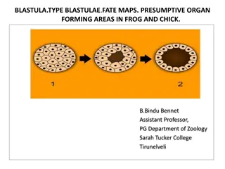
Development of Extraembryonic Membranes in Frog Embryos

The developmental stages of extraembryonic membranes in frog embryos are visually illustrated in a series of images. These images showcase the intricate processes involved in the formation of structures such as the amnion, chorion, yolk sac, and allantois, leading up to the development of the placenta. Detailed depictions of nephrotomal bands, sclerotome, and dermatome are also provided, shedding light on the embryonic development of frogs. Each image captures crucial aspects of embryonic growth, offering valuable insights into the intricate developmental biology of frog embryos.

Uploaded on Sep 20, 2024 | 3 Views
Download Presentation

Please find below an Image/Link to download the presentation.
The content on the website is provided AS IS for your information and personal use only. It may not be sold, licensed, or shared on other websites without obtaining consent from the author.If you encounter any issues during the download, it is possible that the publisher has removed the file from their server.
You are allowed to download the files provided on this website for personal or commercial use, subject to the condition that they are used lawfully. All files are the property of their respective owners.
The content on the website is provided AS IS for your information and personal use only. It may not be sold, licensed, or shared on other websites without obtaining consent from the author.
E N D
Presentation Transcript
B.Bindu Bennet Assistant Professor, PG Department of Zoology Sarah Tucker College Tirunelveli
INTESTINE IN FROG Each lateral plate splits into two layers of mesoderm The parietal or somatic mesoderm The visceral or splanchnic mesoderm The space between these two layers is the coelom.
According to McEwen therefore are originally dorsal extension of the coelom. At this stage the coelom is double i.e one coelom on each side of the embryonic gut.
DEVELOPMENT OF EXTRA EMBRYONIC MEMBRANES
DEVELOPMENT OF AMINON AND CHORION
TYPES OF PLACENTA Four type of placenta : Cotyledonary placenta Zonary placenta Discoidal placenta Diffuse placenta