Developmental Aspects of the Immune System and Innate Defenses Overview
Explore the developmental aspects of the immune system, from stem cells to self-tolerance, and the innate defenses like antiviral proteins, interferon, C-reactive proteins, and more. Understand the immune system's functional system, adaptive defenses characteristics, and the cell-mediated immune response.

Uploaded on | 0 Views
Download Presentation

Please find below an Image/Link to download the presentation.
The content on the website is provided AS IS for your information and personal use only. It may not be sold, licensed, or shared on other websites without obtaining consent from the author. If you encounter any issues during the download, it is possible that the publisher has removed the file from their server.
You are allowed to download the files provided on this website for personal or commercial use, subject to the condition that they are used lawfully. All files are the property of their respective owners.
The content on the website is provided AS IS for your information and personal use only. It may not be sold, licensed, or shared on other websites without obtaining consent from the author.
E N D
Presentation Transcript
Immunity (3) A.I MUSTAFA ALNORRI
Developmental Aspects of the Immune System Stem cells arise from embryologic liver & spleen Self tolerance develops in Thymus (T-cells) & bone marrow (B-cells) Immunocompetence: the library of receptors is genetically determined Immune system degrades with aging
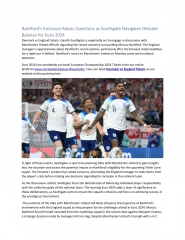
Innate, Internal Defenses Antiviral proteins: interferon & complement Interferon: some cells produce & release interferons (IFNs) when invaded by virus Released interferons stimulate nearby cells to produce proteins (PKR) that interfere with viral replication by disrupting protein synthesis & the ribosome Not virus specific.
Interferon (IFN) Figure 21.5
Innate, Internal Defenses C-reactive proteins (CRP) produced by the liver in response to inflammatory molecules can activate the classical pathway by binding to membrane & activating C1. Also participates in opsonization. Fever a systemic response to infection. Leukocytes & macrophages release pyrogens that raise the hypothalamic set point for temperature
Immune System functional system rather than organ system Hematopoetic Vasculature Lymphatic Fig 21.1
Adaptive Defenses: Characteristics Specificity: directed at specific targets Systemic: not restricted to initial site of infection / invasion Memory: after initial exposure & activation, a more rapid & more vigorous response is made to subsequent exposures to pathogens (secondary response)
Cell Mediated Immune Response T-cell activation: involves recognition of PM surface antigens only Antigen is combined with MHC & displayed on PM T-cell receptors: bind to the MHC & are stimulated by the associated antigen The addition of a co-stimulator (cytokines, interleukins, etc) prompts the T-cell to form a clone In the absence of a co-stimulator the T-cell becomes tolerant to antigen (anergy)
Cell Mediated: MHC MHC occurs as two classes MHC I on virtually all tissue cells MHC II only on PM some immune system cells
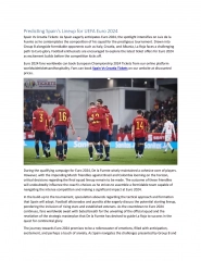
Cell Mediated: MHC display properties Figure 21.16a MHC I on virtually all tissue cells Display only proteins produced inside the cell Endogenous antigens = foreign proteins produced by the cell (viral / cancer) Stimulate the CD8* cell population form cytotoxic T-cells (Killer T, TC) *formerly T8 cells
Cell Mediated: MHC display properties Figure 21.16b MHC II found only on PM of B-cells, some T-cells & APCs Display proteins derived from a phagocytized target Exogenous antigen: foreign protein from outside the cell presented to PM surface Stimulates the CD4* cell population form Helper T-cells (TH) *formerly T4 cells
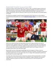
Cell Mediated: T-cell roles Helper T-cells (TH) stimulate B-cells & other T-cells to proliferate Figure 21.18
Cell Mediated: T-cell roles Activated TH cells interact with B-cells displaying antigen & produce cytokines that prompt the B-cell to mature & form antibody Figure 21.18
Cell Mediated: T-cell roles TH cells also produce cytokines that promote TC cells TH cells recruit other WBCs & amplify innate defenses (inflammatory) Subpopulations of TH cells specialize in specific sets of activations Figure 21.18
Cell Mediated: T-cell roles Cytotoxic T-cells (TC, Killer T): directly attack & kill cells with specific antigen Activated TC cells are co-stimulated by TH cells
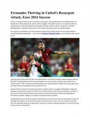
Cell Mediated: T-cell roles Figure 21.19a TC mechanism (Cytotoxic T-cells, Killer T) TC binds to cell & releases perforin & granzymes In the presence of Ca2+ perforin forms pores in target cell PM Granzymes enter through pores & degrade cellular contents TC then detaches & moves on Macrophages clean up
Cell Mediated: T-cell roles Other T-cells *Regulatory T-cells (TReg): release inhibitory cytokines that suppress B-cell & T-cell activity Help to prevent autoimmune events *formerly Suppressor T (TS) Gamma Delta T-cells (Tgd): live in the intestine. Function in surveillance & are triggered much like NK cells
Organ Transplants/Rejections Types of Organ Transplants Autograft: tissue graft from one body site to another (same person) Isograft: graft received from a genetically identical donor (identical twin) Allograft: graft received from genetically non-identical donor (same species) Xenograft: graft received from another species of animal
Organ Transplants/Rejections Transplant rejection: mediated by the immune system (especially TC, NK, antibodies) Auto/Isograft: MHC compatible Xenograft: most MHC incompatible Allograft: attempt to obtain the best MHC match Immunosuppressive therapy: used to delay/prevent rejection Corticosteroids: suppress inflammation Antiproliferative: prevent/kill rapidly dividing cells Immunosuppressant: prevent/kill rapidly dividing cells Side effects tend to be harsh Increased risk of infection
Immunologic Dysfunction Immunodeficiency Congenital/Genetic: varied inborn errors Acquired: Drugs: immunosuppressive / cancer drugs Radiation therapy marrow Cancer: can be viewed as a failure of immune surveillance Hodgkin s disease: lymph node cancer AIDS/HIV: kills TH cells
Immunologic Dysfunction Autoimmune disease: production of antibody & TH against self tissues Examples & tissue effected Multiple sclerosis: white matter of nervous system Graves disease: thyroid Type I diabetes mellitus: beta cells of pancreas Systemic Lupus Erythrematosis: (anti DNA) kidneys, heart, lungs & skin Rheumatoid Arthritis: destroys joints (cartilage) Glomerulonephritis: impaired renal function (may be secondary to other autoimmune disease)
Hypersensitivities: Types Immediate hypersensitivity (Type I): symptoms within seconds of exposure to an allergen (requires sensitization = previous exposure) Figure 21.21
Hypersensitivities: Type I Fast response which occurs in minutes, rather than multiple hours or days. Free antigens cross link the IgE on mast cells and basophils which causes a release of vasoactive biomolecules.Testing can be done via skin test for specific IgE. Mediators: IgE. disorders Anaphylaxis (IgE mediated; mast / basophils) Local: histamine induced vasodilation & increased permeability. Watery eyes, runny nose, itching & redness. Respiratory allergy induced Asthma Atopy:
Hypersensitivities: Types II Antibody (IgM or IgG) binds to antigen on a target cell, which is actually a host cell that is perceived by the immune system as foreign, leading to cellular destruction via the MAC. Testing includes both the direct and indirect Coombs test Mediators: IgG,IgM,complement,MAC disorders rheumatic heart disease (RHD) Autoimmune hemolytic anemia
Hypersensitivities: Types III Antibody (IgG) binds to soluble antigen, forming a circulating immune complex. This is often deposited in the vessel walls of the joints and kidney, initiating a local inflammatory reaction. Mediators:IgG,,complement,neutrophil. Disorders: Rheumatoid arthritis (RA) Systemic lupus erythematosus,
Hypersensitivities: Type IV Helper T cells (specifically Th1 helper t cells) are activated by an antigen presenting cell. When the antigen is presented again in the future, the memory Th1 cells will activate macrophages and cause an inflammatory response. This ultimately can lead to tissue damage. Mediators: T cell Disorders:Contact dermatitis is a type of skin inflammation (dermatitis). Delayed hypersensitivity (cell mediated) takes one to three days to react.
")


")