Diagnostic Imaging Studies and Techniques
This content provides an overview of diagnostic imaging studies and techniques used for various medical conditions. It covers topics such as ordering schemes, X-ray scans, chest, abdominal, urinary, musculoskeletal, and head and neck imaging. The list of symptoms accompanied by recommended imaging studies for accurate diagnosis is also included. Specific body areas and conditions like difficulty speaking, double vision, fever/headache, hemorrhagic stroke, and ischemic stroke are discussed, along with the relevant imaging modalities such as CT scans, MRIs, and contrast imaging. Additionally, imaging studies for conditions like multiple sclerosis, seizures, trauma, severe headaches, enlarged thyroid, lymphadenopathy, and masses are outlined. The content emphasizes the importance of utilizing appropriate imaging techniques for different symptoms and conditions.

Download Presentation

Please find below an Image/Link to download the presentation.
The content on the website is provided AS IS for your information and personal use only. It may not be sold, licensed, or shared on other websites without obtaining consent from the author.If you encounter any issues during the download, it is possible that the publisher has removed the file from their server.
You are allowed to download the files provided on this website for personal or commercial use, subject to the condition that they are used lawfully. All files are the property of their respective owners.
The content on the website is provided AS IS for your information and personal use only. It may not be sold, licensed, or shared on other websites without obtaining consent from the author.
E N D
Presentation Transcript
Dr. Mohamed Abukar Sharif (Dr.MASH)
Part I Ordering schemes Part II X-rays Scan Part III Chest Abdominal Urinaty Musculoskeletal Head and Neck
Part IV Imaging anatomy and pathology Neuroimaging
The following is a list of Pt. symptoms accompanied by the imaging studies that may be beneficial for arriving at an accurate diagnosis.
BODY AREA BODY AREA SYMPTOM/CONDITIO N N DIFFICULTY SPEAKING DOUBLE VISION FEVER/HEADACHE HEMORRHAGIC STROKE ISCHEMIC STROKE SYMPTOM/CONDITIO IMAGING STUDIES IMAGING STUDIES B Brain rain DIFFICULTY SPEAKING DOUBLE VISION FEVER/HEADACHE HEMORRHAGIC STROKE ISCHEMIC STROKE CT SCAN AND /OR CT SCAN AND /OR MRI MRI CONTRST CONTRAST CT SCAN FOLLOWED BY CONTRST MRI CONTRAST CT CT SCAN FOLLOWED BY MRI MRI CT AND/OR AND/OR MRI MRI MRI CT SCAN MAY MISS IN 1 24 PRE/POST CRANIAL CT SCAN PRE/POST CT SCAN MAY MISS IN 1ST 24hr PRE/POST CONTRST CRANIAL MRI CT SCAN PRE/POST CONTRST ST hr MULTIPLE SCLEROSIS SEIZURE TRAUMA SEVERE HEADACHE MULTIPLE SCLEROSIS SEIZURE TRAUMA SEVERE HEADACHE CONTRST MRI MRI MRI CONTRST MRI MRI
BODY AREA NECK BODY AREA NECK SYMPTOM/CONDITION SYMPTOM/CONDITION IMAGING STUDIES IMAGING STUDIES ENLARGED THYROID ENLARGED THYROID NUCLEAR THYROID UPTAKE/SCAN AND ULTRASOUND CT SCAN OR CT(WITH/OUT MRI CT OR NUCLEAR THYROID UPTAKE/SCAN AND ULTRASOUND CT SCAN OR MRI CT(WITH/OUT CONTRST MRI CT OR MRI LYMPHADENOPATHY MASS UNKNOWN ETIOLOGY LYMPHADENOPATHY MASS UNKNOWN ETIOLOGY MRI CONTRST) OR ) OR SALIVARY SALIVARY GALND GALND MASS MASS MRI CHEST CHEST CHEST PAIN COUGH/FEVER/DYSPNEA/W HEEZING HEMOPTYSIS/LUNG NODULE TRAUMA /PLEURAL EFFUSION UNRESOLVING CHEST PAIN COUGH/FEVER/DYSPNEA/W HEEZING HEMOPTYSIS/LUNG NODULE TRAUMA /PLEURAL EFFUSION UNRESOLVING COUGH CXR CXR CXR CT CXR CT SCAN SCAN CHEST CXR SCAN CHEST CHEST CT CXR AND/OR CHEST SCAN CHEST CXR CT SCAN AND/OR CHEST CT SCAN CT COUGH CXR
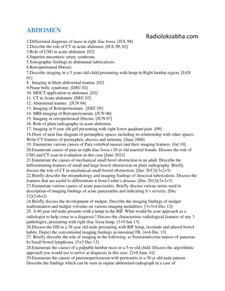
BODY AREA ABDOMEN BODY AREA ABDOMEN ASCITES SYMPTOM/CONDITION SYMPTOM/CONDITION IMAGING IMAGING STUDY STUDY ASCITES CHOLECYSTITIS INTESTINAL OBSTRUCTION CT U/S HIDA SCAN ABDOMINAL WITH BARIUM ENEMA OR U/S ULTRASOUND ABDOMINAL ABDOMINAL X X- -RAY FOLLOWED MRI PELVIC U/S, PELVIC CT SCAN OR U/S U/S HIDA SCAN ABDOMINAL X RAY WITH BARIUM ENEMA OR U/S ULTRASOUND ABDOMINAL CT ABDOMINAL U/S RAY FOLLOWED BY MRI PELVIC U/S, CT.OR PELVIC ULTRASOUND SCAN OR U/S CHOLECYSTITIS INTESTINAL OBSTRUCTION X RAY GALLSTONE TRAUMA APPENDICITIS HIP GALLSTONE TRAUMA APPENDICITIS HIP PAIN CT SCAN U/S SCAN PELVIC PELVIC PAIN BY PELVIC URINE PELVIC PAIN URINE RETENSTION PAIN RETENSTION CT.OR MRI ULTRASOUND MRI
< < HEAD AND NECK MRI SEIZURES MRI BEST BEST MRI=CT HYDROCEPHALUS MRI=CT CT BEST ACUTE HEMORRHAGE CT BEST VASCULAR LESION HEADACHE SCREENING CEREBRAL INFRACTION SALIVARY GLANDS NASOPHARYNX HEAD TRAUMA NEURODEGENERATIVE ACUTE HEADACHE DEMENTIA PITUITARY LESION LARYNX PARANASAL SINUSES THORAX CARDIAC MASSES AORTIC DISSECTION PERICARDIUM LUNG NODULES PERSISTENT PNEUMONIA COPD PULMONARY FIBROSIS MEDIASTINUM HILAR MASS LIVER/SPLEEN PANCREAS/KIDNEYS ABDOMEN HEMANGIOMA VENOUS THROMBOSIS LIVER METASTASIS RENAL TUMORS AORTIC DISEASE ADRENAL/TRAUMA LYMPHADENOPATHY
MRI URETINE FIBROID ENDOMETRIUM PROSTATE CA MRI BEST BEST MRI=CT CERCAL CA RECTAL CA OVARIAN CA BLADDER CA MRI=CT CT BEST ADENOPATHY DIVERTICULITIS APPENDICITIS CT BEST PELVIC MUSCUL- SKELETAL SHOULDER JOINT FRACTURE KNEE/ANKLE/ELBOW JOINT LOOSE BODY SOFT TISSUE NEOPLASM BONE NEOPLASM WRIST/HAND LIGAMENTS/CARTILA GE
Anatomical structures tocheck: 1. Trachea and bronchi 2. Hilar structures 3. Lung zones 4. Pleura 5. Lung lobes and fissures 6. Costophrenic angles 7. Diaphragm 8. Heart 9. Mediastinum 10. Soft tissues 11. Bones 12. Below diaphragm and hidden areas
Check the patient's identity Note the image date and time Note the image projection: Check if a posterioranterior (PA) or anterior posterior (AP) projection was used, and note if the patient was standing, sitting or supine? Was the mobile X-ray machine used? The image annotations are oftenuseful: This is a mobile chest X-ray taken with the patient supine, at 11.25 am in theresuscitation room. The patient's name, ID number and date of birth are annotated. Note the side marker iscorrect.
Metal (bright white) Bone/mineral (white) Fluid/soft tissue (grey) Fat (dark gray) Air (black)
1. Time (reduce to a minimum the time you spend around an X-ray source) 2. Distance(X-ray dose is inversely proportional to the distance squared) 3. Shielding (aprons composed of metal that block x-rays)
X-ray can dislodge electrons from the shell of an atom. This results in the production of an ion (free radical is an unstable molecule with an extra electron. Free radicals become stable by donating their extra electron to other molecules within cells of the body.
This process permanently damages protein, DNA and other vital molecules. Among the ill effects reported from radiation exposure are birth defects, cancer and cataracts.
In case of pneumonia and dehydration order the x-ray of chest


")
")
")