Explore the fascinating world of Optical Coherence Tomography (OCT) imaging to detect various eye diseases and conditions such as Drusen, Retina Detachments, Macular Edema, and more. Understand the anatomy of the eye and how OCT technology helps in early diagnosis and treatment. Delve into the detailed cross-sections and images to enhance your knowledge of ocular health.
Please find below an Image/Link to download the presentation.
The content on the website is provided AS IS for your information and personal use only. It may not be sold, licensed, or shared on other websites without obtaining consent from the author. If you encounter any issues during the download, it is possible that the publisher has removed the file from their server.
You are allowed to download the files provided on this website for personal or commercial use, subject to the condition that they are used lawfully. All files are the property of their respective owners.
The content on the website is provided AS IS for your information and personal use only. It may not be sold, licensed, or shared on other websites without obtaining consent from the author.
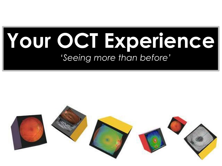
Did you know your OCT experience can help detect many eye diseases ?
DRUSEN Always based at the RPE / Bruchs Complex, Drusen cast no shadows. They tend to be of similar reflectivity as the RPE. They show up at the RPE/BM on the En Face scans.
PED PED Pigment Epithelial Detachments occur when serous fluid lifts the whole Retina from Bruchs membrane. If BM is ever visible separately, this is always pathology. Serous fluid is dark (hypo- reflective) and casts no shadow.
CMO CMO Cystoid Macula Oedema shows generally dark serous fluid in cysts that don t cast shadows. Note in this case there is also serous retinopathy present and some haziness from blood plasma in the larger cystic spaces.
CSR (CSCR) CSR (CSCR) Central Serous Retinopathy (Chorio- Retinopathy) classically affects males in their mid thirties roughly. Notice RPE/BM are intact and the neural retina is lifted by serous fluid which casts no shadow.
ERM ERM Epi Retinal Membranes are extremely common and often don t cause any symptoms. Normally, treatment won t be offered unless the acuity has dropped below 6/24 on average. Traction and Pucker are best viewed with the En Face function.
MACULA HOLE Macula Hole This is a full thickness Macula Hole. Note the reverse shadowing at the central Choroid. This sign is often termed the Kissing Slugs and is not to be mistaken with a lamellar hole or Pseudo hole, often caused by an ERM. (Note the Serous Cysts present here).
WET AMD ERM Epi Retinal Membranes are extremely common and often don t cause any symptoms. Normally, treatment won t be offered unless the acuity has dropped below 6/24 on average. Traction and Pucker are best viewed with the En Face function.
VMT VMT Vitreo-Macular Traction can be a precursor to a Posterior Vitreous Detachment (PVD) which is very common. Sometimes, VMT can pull away some of the Foveal pit where the vitreous is loosely attached. A hyper-reflective piece of Foveal tissue may be seen attached to the Vitreal face, known as an Operculum.
KERATOCONUS (KC) Keratoconus (KC) Here you can make out the Conical cross section of the patients Cornea. It is rare to be able to actually diagnose KC with anterior OCT, but the Pachymetry Maps can also aid with corrected IOPs and the Anterior Chamber Angles can also be measured, for Glaucoma checks.


")