In the realm of DNA labeling and radioactive decay, explore the composition of atoms, the concept of isotopes, and the three types of decay processes - alpha decay, beta (-) decay, and beta (+) decay. Uncover the dynamics of nucleus degradation, transmission between elements, radiation emission, and elemental transformation. Delve into the fundamental principles governing atomic structures and the forces that bind nuclei together versus those that lead to radioactive instability.
Please find below an Image/Link to download the presentation.
The content on the website is provided AS IS for your information and personal use only. It may not be sold, licensed, or shared on other websites without obtaining consent from the author. If you encounter any issues during the download, it is possible that the publisher has removed the file from their server.
You are allowed to download the files provided on this website for personal or commercial use, subject to the condition that they are used lawfully. All files are the property of their respective owners.
The content on the website is provided AS IS for your information and personal use only. It may not be sold, licensed, or shared on other websites without obtaining consent from the author.
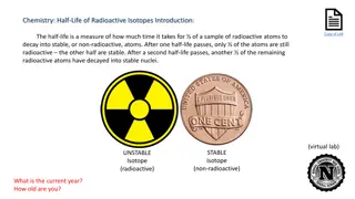
An atom is composed of positively charged nucleus that is surrounded by negatively charged electrons. The number of electrons is equal to the number of protons = Atomic Number Sum of protons and neutrons present in the nucleus = Atomic mass Strong nuclear forces holds the positive protons and neutral neutrons together in the nucleus. In stable nuclei, nuclear force is strong enough to hold the nucleus permanently. Unstable nuclei (either have too many neutrons or too many protons): These unstable nuclei balance themselves by giving off the excess protons or neutrons. This is called radioactive decay. Unstable nuclei are radioactive and emits radiation.
Atoms that have the same amount of protons but differs in neutrons amount are called isotopes. For example, carbon-12, carbon-13, and carbon-14 are three isotopes of the element carbon with mass numbers 12, 13, and 14, respectively. The atomic number of carbon is 6, which means that every carbon atom has 6 protons so that the neutron numbers of these isotopes are 6, 7, and 8 respectively. The spontaneous degradation of nucleus & transmission of one element to another with consequent emission of rays ( or ) particles is known as radioactivity.
Three types of Decay: 1. Alpha ( ) Decay or alpha radiation : Release of particle (2He4) Parent nucleus Daughter nucleus + alpha particle An alpha particle is identical to the nucleus of a normal Helium atom i.e., doubly ionized helium atom (He2+) When an atom emits an alpha particle in alpha decay, the atom's mass number decreases by four . The atomic number of the atom goes down by two, as a result of the loss of two protons the atom becomes a new element.
2. Beta (-) Decay or beta radiation (too many neutrons) + e - + + + Lost high energy electron is called particle. Atomic mass will be same, however, atomic number will be enhanced by one. New element is formed; one place higher in the periodic table.
3. Beta (+) Decay or positron radiation (too many protons) + e + + + + + + + + Beta-plus decay (also called positron emission) occurs when a proton is converted to a neutron, and in the process, emits a positively charged electron (a positron). A positron is a particle identical to an electron except that it has a positive charge Atomic number decreases by one and mass number remains the same . One place lower in the periodic table.
Gamma Radiation A gamma ray is a high-energy photon emitted by a radioisotope. Often are emitted along with alpha and beta particles. Gamma radiation doesn t have a positive or negative charge. Gamma rays are similar to X-rays, but they have even greater energy. Gamma radiation can only be stopped by a thick layer of lead or concrete. Other examples:
Separating Alpha, Beta, and Gamma Particles >Alpha and beta particles are deflected in opposite directions- alpha particles toward the negative plate and beta particles toward the positive plate. >Gamma rays are un-deflected. Path of the beta particle is curved more than the alpha. This is due to the mass.
Unstable or Stable nuclei: What type of decay likely to occur? If a radioisotope lies left or right of stability line, it is unstable and likely to decay to become stable Radioisotopes left to stability line have too many neutrons and they are likely to undergo beta- decay. Radioisotopes right to stability line have Too many protons and they are likely To undergo beta+ decay. High number of protons (> 82): alpha decay
Stable & Unstable isotopes Unstable isotopes: Spontaneously fall apart, emitting particles and energy (radioactivity) : Radioisotopes Stable isotopes: Remain as they are indefinitely Hydrogen =1H,2H stable, 3H unstable Carbon: 12 C, 13 C stable, 14 C unstable Oxygen: 16 O, 18 O stable, 17 O unstable Deuterium H-2, an isotope twice as heavy as hydrogen, is predominantly used in nutrition research, Nitrogen-15 is the most common stable isotope used in agriculture. 13 N, 12 N: + Release 16 N, 17 N, 18 N, 19 N, 20 N, 21N ..: - Release
APPLICATION OF RADIOISOTOPES IN BIOLOGICAL SCIENCES 1. Radioisotopes are frequently used for tracing metabolic path ways 2. Labeling of nucleic acids 3. Study of the mechanism & rate of absorption , accumulation & translocation of inorganic & organic compounds in the animal and plants. 4. Radiolabeled drugs are useful in pharmacokinetic studies (site of accumulation, rate of accumulation, rate of metabolism & metabolic products ). 5. Virtually any enzyme reaction can be assayed using radioactive tracer methods. 6. Radioisotopes have been used in study of the mechanism of enzyme action and also in studies of ligand binding to membrane receptors. 7. Radioimmuno assays are useful in analysis of hormones, growth factors, tumour markers, cytokines, bacterial antigens, vitamin D & various biological molecules. 8. Radioisotopes have role in management of malignancies. Tumour tissues are attacked by beam of radiation. 131I is used for treatment of thyroid cancer. 60Co is the source of radiation.
The SI unit of radioactive activity is the becquerel (Bq), in honor of the scientist Henri Becquerel. One Bq is defined as one transformation, decay, or disintegration per second. Since sensible sizes of radioactive material contain many atoms, a Bq is a tiny measure of activity; amounts giving activities on the order of GBq (gigabecquerel, 1 x 10⁹decays per second) or TBq (terabecquerel, 1 x 10¹²decays per second) are commonly used. Another unit of radioactivity is the curie, Ci, it is equal, by definition, to the activity of any radionuclide decaying with a disintegration rate of 3.7 1010Bq, so that 1 curie (Ci) = 3.7 1010Bq. Counts per minute (cpm) is a measure of radioactivity. It is the number of atoms in a given quantity of radioactive material that are detected to have decayed in one minute. Disintegrations per minute (dpm) is also a measure of radioactivity.
Radioactivity measurement: Geiger-Muller counter (G-M Counter) The Geiger Muller counter (Gas based counter) is an instrument used for measuring ionizing radiation. It detects ionizing radiation such as alpha particles, beta particles and gamma rays using the ionization effect produced in a Geiger M ller tube. It is one of the best-known radiation detection instruments Readout: Count per second Absorbed dose
Labeling and Detection of Nucleic acid In molecular biology, hybridization (is a phenomenon in which single-stranded deoxyribonucleic acid (DNA) molecules anneal to complementary DNA or RNA. or ribonucleic acid (RNA) Though a double-stranded DNA sequence is generally stable under physiological conditions, changing these conditions in the laboratory (generally by raising the surrounding temperature) will cause the molecules to separate into single strands. These strands are complementary to each other but may also be complementary to other sequences present in their surroundings. Lowering the surrounding temperature allows the single-stranded molecules to anneal or hybridize to each other. Nucleic acid hybrids can be formed between two strands of DNA, two strands of RNA or one strand of DNA and one of RNA.
Nucleic acid hybridization with a labeled probe is the only way to detect a complementary target sequence in a complex nucleic acid mixture. Nucleic acid probes are oligonucleotides or polynucleotides that can bind with high specificity to complementary sequences. Probes can be complementary to either DNA or RNA and can be from as few as 20 nt to hundreds of nt long. DNA probes RNA probes Oligonucleotide probes
Oligonucleotide probes are short stretches of single-stranded DNA or RNA used to detect the presence of complementary nucleic acid sequences (target sequences) by hybridization. In molecular biology, a nucleic acid probe is a fragment of DNA or RNA which can be radioactively or fluorescently labeled. These probes can be used of nucleotide sequences in are complementary to the sequence in the probe. to detect RNA the or presence DNA analyzed that The under alkaline conditions such as exposure to sodium hydroxide) into single stranded DNA (ssDNA) and then hybridized to the target ssDNA (Southern blotting) or RNA (northern blotting) immobilized on a membrane or in situ. labeled probe is first denatured (by heating or
DNA probes DS OR SS RNA probes SS Oligonucleotide probes SS 1. Heterologous Probe: Probe that is similar, but not exactly the same Mouse probe can be used to search a human genomic library. 2. Homologous: Probe that is exactly complementary to the nucleic acid sequence of interest
Label Location There are two ways to label a DNA molecule by the ends (end labeling) OR all along the molecule (uniform labeling) Uniform labeling: A. Nick translation The endonuclease DNase I is used to create nicks at random sites in the strand of double stranded target DNA DNA polymerase I is used to add nucleotide residues to the free 3 -hydroxyl ends created during the DNase I nicking process. As the DNA polymerase I extends the 3 -end, the 5 - to 3 exonulecase activity of the enzyme simultaneously removes bases from the 5 -end of the nick. Sequential addition of bases on to 3 -end with the simultaneous removal of bases from the 5 -end results in translation of the nick Along the DNA molecule. DNA ligase
Pancreatic DNase I E. coli DNA polymerase I DNA ligase When performed in the presence of a radioactive ([ -32P]dNTP the newly synthesized strand becomes radioactivity labeled.
B. Random priming: This is an alternative method for preparing uniformly labeled DNA is by oligo-nucleotide primed DNA synthesis with hexanucleotide (or longer oligomers) of random sequences The klenow fragment is used as this enzyme lacks the 5 -3 exonuclease activity of DNA Polymerase I. It fills gaps between adjacent primers. Labeled nucleotides are incorporated Into new DNA that is synthesized.
MCQ Nucleic acids are readily labeled with tags that facilitate detection or purification. Which of the following components are required for the 3 - end labeling of DNA with radioactive phosphorous? (A) Terminal deoxynucleotidyl transferase and (gamma) 32P dNTP (B) Polynucleotide kinase and (gamma) 32P dNTP (C) Terminal deoxynucleotidyl transferase and (alpha) 32P dNTP (D) Polynucleotide kinase and (alpha) 32P dNTP


 Decay or beta radiation")
 Decay or positron radiation")


,")