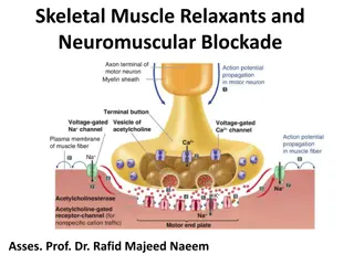
Dose-Dependent Association Between Intermediate-Acting Neuromuscular Agents and Postoperative Respiratory Complications
Anesthesia involving intermediate-acting neuromuscular-blocking agents (NMBAs) may lead to postoperative respiratory issues. This study explores the dose-dependent relationship and the impact of neostigmine reversal. Patients over 18 undergoing surgery with tracheal intubation and extubation, excluding recent surgeries, were evaluated for respiratory complications. The primary outcome included respiratory failure, pulmonary edema, tracheal reintubation, and pneumonia within the first 3 days post-extubation. Of 48,499 cases analyzed, 3.7% experienced respiratory complications, with pulmonary edema being the most common.

Download Presentation

Please find below an Image/Link to download the presentation.
The content on the website is provided AS IS for your information and personal use only. It may not be sold, licensed, or shared on other websites without obtaining consent from the author.If you encounter any issues during the download, it is possible that the publisher has removed the file from their server.
You are allowed to download the files provided on this website for personal or commercial use, subject to the condition that they are used lawfully. All files are the property of their respective owners.
The content on the website is provided AS IS for your information and personal use only. It may not be sold, licensed, or shared on other websites without obtaining consent from the author.
E N D
Presentation Transcript
RESPIRATORY MANAGEMENT
Contents Anatomy and physiology Anatomy and physiology Tracheostomy Tracheostomy Cuff management Cuff management Passy Muir Passy Muir Valve Invasive ventilation Invasive ventilation Non Non- -invasive ventilation invasive ventilation Fisher & Fisher & Paykell Paykell Humidification Cough Cough assistantor assistantor CPAP CPAP Blood Gas Blood Gas Valve Humidification
What is a tracheostomy? Surgical opening in anterior wall of trachea Provides airway access into the trachea Bypasses an upper airway obstruction Facilitates mechanical ventilation and weaning in long term intubated patients.
Indications for tracheostomy Upper airway obstruction Assistance in secretion clearance Pronged airway protection Assistance in artificial ventilation
Components of a tracheostomy Tracheal dilator---used for insertion of trache into stoma Outer cannula Inner cannula Flange---side pieces, flexible or rigid, the size and type printed on Size---size is recorded, Tacoe Twist & Portex Blue Line
Components of a tracheostomy Cuff---balloon surrounding outer cannula Pilot balloon---balloon on the outside, attached to the cuff via thin tube Fenestrations---small holes in cannula of trachea Cap---white or red cap sits on hub of inner cannula Bed site tracheostomy emergency box
Types of tracheostomies Single lumen 1. Outer tube only, no inner 2. Usually first sited 3. Non-fenestrated Double lumen 1. Inner and outer cannula 2. Trends to be longer term 3. May or may not be fenestrated
Types of tracheostomies Non-fenestrated 1. No holes in the cannula, is usually the first trache sited Fenestrated 1. An opening or hole in the posterior wall of the trache 2. Trends be single hole or clover leaf pattern 3. Cuff deflated assists airflow up into the larynx for speech 4. When suctioning, inner tune must be changed to a non- fenestrated tube to prevent tracheal trauma
Types of tracheostomies Cuffed tubes 1. Cuff maintains a seal against the tracheal wall 2. Minimizes the movement of the tube therefore reduces the risk of tracheal trauma, oesophagus and blood vessel erosion 3. Minimizes but not necessarily prevents aspiration 4. Allow mechanical ventilation to occur Non-cuffed tubes 1. Useful in the weaning process 2. May allow the patient speak
Suctioning a tracheostomy Aim: is to stimulate a cough and remove excess secretions Sizing the catheter: 1. Catheter is no more than half of trache lumen 2. Divide trache size by 2 and multiple by 3 (ie. Shiley size 8. Req: 8/2x3=12 cathter
Technique for suctioning Do not suction with fenestrated inner tube as risk of trauma Carefully insert catheter into tracheostomy until cough and gentle resistance felt Withdrawl catheter half cm, apply continuous suction (no twirling, no twisting) upon exiting
Cuff Care Cuff maintains seal against tracheal wall to allow for ventilation and minimize aspiration without tracheal ischemia and trauma. Pilot balloon represents the cuff. Aim for minimal occlusion pressure Need to check cuff pressure each shift and record measurements
Synchronised suction/cuff deflation Two persons procedure: Suction mouth and/or pharyngeal area first Where one person lets the cuff down whilst the other person simultaneously suctions This allows the secretions sitting above the cuff to be removed rather than aspirated when the cuff is deflated
Passy Muir Valve One way speaking valve If using a non-fenestrated tube, the cuff must be DEFLATED and the inner cannula should be removed if not fenestrated If fenestrated, it is possible to keep the cuff inflated and create a voice, although this is not routinely recommended If humidified oxygen or air is required, this can be placed over the speaking valve in the normal manner Instruct patient to breathe in via the tracheal tube, and blow out through the mouth If voice sounds wet or gurgly , ask to cough and clear secretions. If difficulty, tracheal suctioning might be helpful Monitor carefully. Remove the speaking valve if necessary. Always refer to SLT for advice Attaching the speaking valve to ventilator circuit
Ventilators Types of ventilators:Nippy 3+, Nippy 4, Trilogy, etc. Mode (PSV, PCV and CPAP) Settings (IPAP, EPAP, Ti, Backup rate) Indications for adjusting the settings and alarms Importance of exhalation valve SenTec monitoring (indications & normal range) Circuit single circuit (HME) and F&P circuit
Non-invasive ventilation Indications Components Types of face masks Humidification Oxygen therapy
Fisher & Paykell Humidifier Fisher & Paykell machine Circuit
Cough Assistor Is a non-invasive therapy that safely and consistently removes secretions in patients with an effective ability to cough. Clears secretions by gradually applying a positive pressure to the airway, then rapidly shifting to negative pressure. The rapid shift in pressure produces a high expiratory flow, stimulating a natural cough.
Benefits of Cough Assistor Removes secretions from the lungs Reduces the occurrence of respiratory infections Safe, non-invasive alternative to suctioning Easy for patients and staff to operate
CPAP CPAP is maintenance of positive airway pressure greater than ambient pressure during inspiration and expiration. It may be given via a mask, mouthpiece, endotracheal tube or tracheostomy tube. The aim is to improve gas exchange and improve the work of breathing. It may be used in the following circumstances: Respiratory failure where the patient is able to breathe spontaneously and maintain airway Method of weaning a patient from a ventilator Supportive measure where mechanical ventilation not considered appropriate or not acceptable to patient Acute respiratory distress (ARDS) Following major abdominal or thoracic surgery to prevent atelectasis Pulmonary oedema Sleep apnoea Some patients require ventilator settings to be altered to CPAP mode as part of a weaning programme and vice versa.
Blood Gas Normal Values pH: (7.34-7.45) PCO2: (35-45mmHg / 4.67-6.00kPa) PO2: (80-105mmHg / 10.7-14kPa) HCO3:(22-26mmol/L) Base Excess (BE): (-2--+3mmol/L)
Ventilation PaCO2 lungs eliminate respiratory acid as carbon dioxide (CO2) When breathing effectively and moving adequate volumes of air in and out of the lungs PaCO2 will stay within normal limits Other scenarios are: Respiratory acidosis pH less than 7.35 and PaCO2 greater than 6kPa Respiratory alkalosis pH greater than 7.45 and PaCO2 less than 4.67KPa
Respiratory acidosis Hypoventilation, less air will be moved in and out of lungs, causing CO2 to be retained. May be caused by: Airway obstruction Impaired alveolar filling e.g. bronchopneumonia Depression of respiratory centre in brain e.g. drug overdose Decreases force of cardiac contractions
Respiratory alkalosis Hyperventilation moves large amount of air in and out of lungs Respiratory acid load reduces causing an excess of alkali May be caused by: Fall in oxygen levels e.g. lung disease, severe anaemia Stimulation of central nervous system causing raised intracranial pressure e.g. head injury, tumour Can interfere with tissue oxygenation