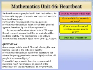
E-Rate Update FY 2019-2020
Sandhills CTO Meeting E-Rate Update for FY 2019-2020 provides detailed information on application filing, planning steps, tariff impacts, and contact details for further queries. Stay informed to meet the submission deadlines and procurement requirements.

Download Presentation

Please find below an Image/Link to download the presentation.
The content on the website is provided AS IS for your information and personal use only. It may not be sold, licensed, or shared on other websites without obtaining consent from the author.If you encounter any issues during the download, it is possible that the publisher has removed the file from their server.
You are allowed to download the files provided on this website for personal or commercial use, subject to the condition that they are used lawfully. All files are the property of their respective owners.
The content on the website is provided AS IS for your information and personal use only. It may not be sold, licensed, or shared on other websites without obtaining consent from the author.
E N D
Presentation Transcript
ADVANCED CARDIAC LIFE SUPPORT(ACLS) Dr. Samad Shams Vahdati, MD Professor of emergency medicine
ADVANCED CARDIAC LIFE SUPPORT Advanced cardiac life support or advancedcardiovascular life support (ACLS) refers to a set of clinical interventions for the urgent treatment of cardiac arrest and other life-threatening medical emergencies, as well as the knowledge and skills to deploy those interventions.
ACLS is a series of evidence based responses simple enough to be committed to memory and recall under moments of stress. AMERICAN HEART ASSOCIATION (AHA) protocols are considered to be the GOLD standard ACLS protocols It gets reviewed every 5 year, now latest advancements in ecgguidelines.health.org
IMPORTANCE OF BLS IN ACLS ACLS is built heavily upon the foundation of BLS
AHA Adult Chain ofSurvival 1.Immediate recognition of cardiac arrest and activation of the emergency response system 2.Early CPR with an emphasis on chest compressions 3. Rapid defibrillation 4. Effective advanced life support 5. Integrated post cardiac arrest care
AHA PEDIATRIC Chain of Survival
COMPONENT OF HIGH QUALITY CPR IN BLS Scene safety: 1. Make sure the environment is safe for rescuers and victim Recognition of cardiac arrest: 1. Check for responsiveness 2. No breathing or only gasping ( ie, no normal breathing) 3. No definite pulse felt within 10 secs ( Carotid or femoral pulse) 4. (Breathing and pulse check can be performed simultaneously within 10 secs)
Activation of emergency response system: If alone with no mobile phone, leave the victim to activate the emergency response system and get the AED before beginning CPR Otherwise, send someone and begin CPR immediately; use the AED as soon as it is available
WITNESSED VS UNWITNESSED WITNESSED IFALONE ACTIVATE EMS THEN CPR IF 2 RESCUERS START CPR SECOND ONE ACTIVATEEMS UNWITNESSED STARTCPR GIVE FOR 2 MINS ACTIVATE EMS
Chest compression- Adult- 30:2 Children or infant- 30:2 if one rescuer 15:2 if more than one rescuer Compression rate: 100-120/ min Compression depth: Adult- at least 5 cm Children or infant- at least 1/3rd AP diameter of chest
Hand placement: Adult - 2 hands on the lower half of the sternum Children 1 or 2 hands on the lower half of the sternum Infants 2 fingers or 2 thumb defending of the number of rescuers Chest recoil: allow full recoil of chest after each compression; do not lean on the chest after each compression. Minimizing interruption: Limit interruptions in chest compressions to less than 10 secs.
Adult advanced cardiovascular life support
Shockable VT VF Monomorphic or polymorphic Fine or Coarse VF
Ventricular tachycardia .R-R interval usually regular, not always QRS not preceded by p wave. Wide and bizzare QRS. Difficult to find seperation between QRS and T wave Rate=100-250bpm
Torsades de Pointes Ttwisting of points, is a distinctive form of polymorphicventricular tachycardia characterized by a gradual change in the amplitude and twisting of the QRS complexes around the isoelectricline. Rate cannot be determined.
Ventricular fibrillation A severely abnormal heart rhythm (arrhythmia) thatcan be life-threatening. No identifiable P, QRS or T wave Emergency- requires Basic Life Support Rate cannot be discerned, rhythmunorganized
Unshockable PEA- pulseless electrical activity or EMD- electromechanical dissociation Asystole
Asystole a state of no cardiac electrical activity, hence no contractions of the myocardium and no cardiac output or blood flow. Rate, rhythm, p and QRS are absent
Pulseless electrical activity Pulseless electrical activity (PEA) unresponsiveness and no palpable pulse some organized cardiac electrical activity. previously referred to as electromechanical dissociation
Deliver singledefibrillitor shock CPR-2 mins Checkrhythm Deliver single shock- ifVT /VF persist---CPR 2 mins and give EPINEPHRINE 1 mg Continue CPR 2min Amiodarone/ Lidocaine/ Magnesiumsulfate Vt/ vf Defibrillate: Drug---Shock---Drug---- Shock
Asystole/PEA Identify and RX reversible causes Continue CPR if asystole/PEA Continue CPR (Intubate and establish IV access)
Treatable Causes of Cardiac Arrest: The H s and T s T s H s Hypoxia Hypovolemia Hydrogen ion(acidosis) Hypo-/hyperkalemia Hypothermia Toxins Tamponade (cardiac) Tension pneumothorax Thrombosis, pulmonary Thrombosis, coronary
Defibrillation Biphasic wave form: 120- 200 J Monophasic wave form: 360 J AED- device specific Failure of a single adequate shock to restore a pulse should be followed by continued CPR and second shock delivered after five cycles of CPR
HOW TO USE DEFIBRILLATOR SAFETY If patient not intubated remove o2 delivery devices If intubated either leave bag valve resuscitator attached to Et or remove it If available use self adhesive defibrillation pads Do not place over pacemakers Remove transdermal patches.
PROCEDURE Place sternal paddle over right of the sternum below clavicle Place apical paddle in mid axillary line in 5th IC space Switch on the defibrillator Charge the defibrillator to 200J or 360J Warn all other rescuers to stand clear- ARE YOU CLEAR Visually check all are clear Ensure yourself you are not touching patient or bed I AM CLEAR
Deliver shock Restart cpr with out checking pulse.
Automatic External Defibrillator Switch onAED. Attach electrode pads. Place electrodes as that of manual one Follow voice commands Make sure no one in contact with patient Push shock button.
1-Shock Protocol Versus 3- Shock Sequence Evidence from 2 well-conducted pre/post design studies suggested significant survival benefit with the single shock defibrillation protocol compared with 3-stacked-shock protocols If 1 shock fails to eliminate VF, the incremental benefit of another shock is low, and resumption of CPR is likely to confer a greater value than another shock
Airway and Ventilations Opening airway Head tilt, chin lift or jaw thrust, in addition explore the airway for foreign bodies, dentures and remove them.
Breathing devices BASICAIRWAYS Oropharyngeal airway Nasopharyngeal airway ADVANCED Endotracheal tube Laryngeal mask airway Laryngeal tube Esophageal tracheal tube
commonly 67 mm in an adult female and 78 mm for an adult male
90-115cm 105-130 122-155