Embryo Development and Fertilization in Animal Science
In this comprehensive study, various aspects of embryo development and fertilization in animals are discussed, including errors in fertilization such as polyspermy and polyandry, as well as oocyte development and fertilization processes. The content also explores the stages of embryo development in bovines, highlighting key milestones from zygote formation to blastocyst development.

Download Presentation

Please find below an Image/Link to download the presentation.
The content on the website is provided AS IS for your information and personal use only. It may not be sold, licensed, or shared on other websites without obtaining consent from the author.If you encounter any issues during the download, it is possible that the publisher has removed the file from their server.
You are allowed to download the files provided on this website for personal or commercial use, subject to the condition that they are used lawfully. All files are the property of their respective owners.
The content on the website is provided AS IS for your information and personal use only. It may not be sold, licensed, or shared on other websites without obtaining consent from the author.
E N D
Presentation Transcript
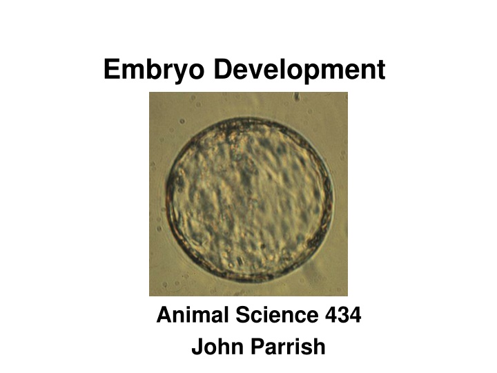
Embryo Development Animal Science 434 John Parrish
Errors in Fertilization Polyspermy - polyandry Multiple sperm penetration Invertebrates excess sperm eliminated because sperm centriole contributes to first embryonic cleavage spindle Mammals Sperm centriole not essential so development continues but fails early to midpregnancy due to multiploidy Occurs most often in aged oocytes due to failure of zona block to polyspermy
Errors in Fertilization (cont.) Polygyny Multiple maternal pronuclei + 1 paternal pronuclei Artificially created only Suppress extrusion of the PBII Androgenote Union of 2 paternal pronuclei Artificially created only From pronuclear exchange
Errors in Fertilization (cont.) Gynogenote Union of 2 maternal pronuclei Artificially created Induced oocyte activation and supression of PBII extrusion Parthenogenesis Activation of the oocyte without a sperm Embryo is either haploid or gynogenesis occurs to form diploid Platties - sperm activates but then gynogenesis occurs and sperm extruded from embryo
Oocyte Development and Fertilization LH Surge (0 hr) Primary Oocyte GVBD (8 hr) Metaphase I MPF MPF GV-Intact PB-1 Ovulation (29 hr) MPF MPF Metaphase II (21 hr) Secondary Oocyte
Zona Pellucida Oocyte Ca2+ Sperm Penetration of the Zona Pellucida and Fusion with the Oocyte (30 hr) Perivitelline Space
Embryo Development in the Bovine 4 cell (75 hr, day 3) 8 cell (90 hr, day 3) 2 cell (62 hr, day 2) 16 cell (120 hr, day 4) Zygote (34 hr, day 1) Hatched Blastocyst (day 9-11) Trophectoderm 32 cell Morula (day 5-6) Blastocoel Compaction Early Blastocyst (day 7-8) Inner Cell Mass Tight Morula (day 6-7) Expanded Blastocyst (day 8-10) Blastocyst (day 7-9)
Fertilization to Cleavage Zygote Zona Pellucida Perivitelline Space Pronuclei Polar Body Blastomere
Fertilization to Cleavage Imprinting Maternal Gene Control Long Cell Cycle
Imprinting Sperm Pronucleus Egg Pronucleus
Imprinting Sperm Pronucleus Egg Pronucleus Androgenote Gynogenote
Imprinting Sperm Pronucleus Egg Pronucleus Controls Androgenote Gynogenote
Imprinting Androgenote Gynogenote Controls Normal Fetus Normal Placenta Normal Pregnancy No Inner Cell Mass Normal Placenta Fails during embryo development Normal Fetus Small Placenta Fails Midpregnancy Maternal and Paternal Genomes Are Expressed Differently in the Embryo and Fetus
Gene Control of Development Embryonic Gene Control Maternal Gene Control Oocyte Growth LH Fertilization Cleavage Surge Transciption Translation No transcription Translation Post-Translation Translation Post-Translation Transcription Translation Post -Translation
Fertilization to Cleavage Maternal Gene Control
Fertilization to Cleavage Long Cell Cycle Penetration to Cleavage 32 hour (Bovine)
Precompaction Cleavage Cell size decreases Cell cycle Embryonic gene control Asynchrony of cell divisions Movement into Uterus Early pregnancy factor
Precompaction Cleavage Cell size decreases Cell cycle Asynchrony of cell divisions Embryonic gene control Movement into Uterus Early pregnancy factor
Precompaction Cleavage 32 hours 13 hours Cell size decreases Cell cycle Embryonic gene control 15 hours Asynchrony of cell divisions Movement into Uterus Early pregnancy factor 30 hours
Cell Cycle Lengths 1st Cell Cycle (zygote 2 cell) 2nd Cell Cycle (2 cell 4 cell) 8 hr <1 hr 8 hr 16 hr 8 hr 2 hr G1 S G2 + M Total = 32 hr Total = 13 hr
Precompaction Cleavage 32 hours Short G1 and G2 13 hours Cell size decreases Cell cycle Short G1 and G2 Embryonic gene control 15 hours Asynchrony of cell divisions Movement into Uterus Early pregnancy factor 30 hours
Precompaction Cleavage Cell size decreases Faster dividing blastomeres go to center of embryo Cell cycle Asynchrony of cell divisions Embryonic gene control Movement into Uterus Early pregnancy factor
Asynchronous Cleavage - Inside Outside Theory Inner Cell Mass If a marked blastomere is placed into the interior of a 8-cell embryo, it and its progeny become part of the ICM. Trophectoderm If a marked blastomere is placed on the outside of a 8-cell embryo, it and its progeny become part of the trophectoderm.
Asynchronous Cleavage Use Create embryos from different species Placenta from one species Host mother Embryo from some other species Donor mother
Precompaction Cleavage Cell size decreases Cell cycle Asynchrony of cell divisions Embryonic gene control Movement into Uterus Early pregnancy factor
Gene Control of Development Embryonic Gene Control Maternal Gene Control Oocyte Growth LH Fertilization Cleavage Surge Transciption Translation No transcription Translation Post-Translation Translation Post-Translation Transcription Translation Post -Translation
Transition from Maternal to Embryonic Gene Control Transcription of the embryonic genome Species First begins Development is dependent on Mouse 1 cell 2 cell Rabbit 2 cell 8 cell Pig 4 cell 8 cell Cattle 4 cell 8-16 cell Sheep 8 cell 16 cell Human 4 cell 8 cell In vitro blocks to development often occur here!!!!!
Precompaction Cleavage 32 hours 13 hours Cell size decreases Cell cycle Asynchrony of cell divisions 15 hours Embryonic gene control Movement into Uterus Early pregnancy factor 30 hours Embryo runs out of key factors coded for by maternal mRNA Cell Cycle Length Increases Pause in G1
Precompaction Cleavage Cell size decreases Cell cycle Asynchrony of cell divisions Embryonic gene control Movement into Uterus Early pregnancy factor
Movement into the Uterus Ampulla Uterine Horn Isthmus < 8 cell > 8 cell Occurs around day 4 Cause Change in estrogen progesterone
Precompaction Cleavage Cell size decreases Cell cycle Asynchrony of cell divisions Embryonic gene control Movement into Uterus Early pregnancy factor
Early Pregnancy Factor Found at 24 - 72 hours after fertilization Mice, hamster, sheep, cattle, swine, human Seen only in viable pregnancy More recent experience in cattle may not agree with this Function Sensitize the uterus to implantation Basis for early pregnancy kit in cattle
Morula to Blastocyst Polarization Compaction
Polarization Polar Blastomeres Microvilli Non-polar Blastomeres
Polarization (cont.) Gap Junctions Tight Junctions
Cell Linage Polar Cells Non-polar Cells 2 polar cells 1 polar 1 non-polar 2 non-polar
Compaction Occurs at fixed time after fertilization Membranes are very close and begin to flatten. Resulting in loss of the round cell outlines. Differentiational event Genome controlled and involves microtubules and microfilaments.
Blastocyst Formation and Hatching Blastocoel formation Hatching
Blastocoel Formation Morula H2O Na+ Gap Junctions Tight Junctions
Blastocoel Formation Morula H2O Na+ Gap Junctions Tight Junctions
Blastocoel Formation Blastocoel Early Morula Blastocyst H2O Na+ Gap Junctions Tight Junctions
Blastocyst Formation and Hatching Blastocoel formation Hatching Early Blastocyst H2O Blastocoel formation not dependent on: Cell number Cell division Embryonic genome expression required Na+ Blastocyst Inner Cell Mass Expanded Blastocyst Trophectoderm
Blastocyst Formation and Hatching Blastocoel formation Hatching Early Blastocyst Enzymatic digestion of zona Plasminogen and plasminogen activator made by embryo Softening of zona by uterine enzymes Increase in size of blastocyst due to water pumping Most important Day 9 - 11 in cattle, 6 in swine, and day 7 - 8 in horses or sheep Blastocyst Expanded Blastocyst Hatching Blastocyst
Formation of Twins Dizygotic Not identical Double ovulation Monozygotic Identical Several potential mechanisms
Formation of Monozygotic Twins Siamese Twins


")
")


")