Embryology and Fetal Development: From Fertilization to Fetal Growth
Explore the fascinating journey of embryology and fetal development, starting from fertilization to the formation of zygote, morula, and blastocysts. Understand the stages of fetal growth and development in the early weeks after conception, including the rapid growth, formation of the embryonic plate, development of the central nervous system, heartbeat initiation, and skin coverage. Dive into the wonders of human life unfolding in the womb.

Download Presentation

Please find below an Image/Link to download the presentation.
The content on the website is provided AS IS for your information and personal use only. It may not be sold, licensed, or shared on other websites without obtaining consent from the author. If you encounter any issues during the download, it is possible that the publisher has removed the file from their server.
You are allowed to download the files provided on this website for personal or commercial use, subject to the condition that they are used lawfully. All files are the property of their respective owners.
The content on the website is provided AS IS for your information and personal use only. It may not be sold, licensed, or shared on other websites without obtaining consent from the author.
E N D
Presentation Transcript
Fertilization Fertilization is also called conception/impregnation/fecundation. Is the fusion of the sperm with the ovum, to form the zygote -The prerequisites for fertilization includes: - Adequate and matured sperm. Ability of sperm to reach and penetrate the zona pellucida and cell membrane of the ovum. Matured ovum. Patent genitalia. 2
The fertilized cell continues its passage through the uterine tube & reaches the uterus 3 - 4 days later. During this time segmentation or cell division continues. Implantation is normally complete by the 11th day after ovulation. The fertilized ovum is called a zygote till it will be 3 weeks after conception. After 3 wks up to 8 wks we call it an embryo. After 8 weeks up to delivery we call it a fetus which when born will be called a baby. 3
The Fetal Development 1. The zygote: - The zygote is the beginning of the embryo. After the zygote formation, typical mitotic division of the segmentation nucleus occur producing two blastomeres. The blastomeres continue to divide by binary division through 4, 8, 16 cells stage until a cluster of cells called Morula is formed. 2. The Morula: - The Morula after spending about 3 days in the uterine tube enters the uterine cavity through the narrow uterine ostium (1mm) on the fourth day in the 16 - 64 cell stage. The transportation is controlled by muscular contraction and cilia in the fallopian tubes. The central cell of the Morula is known as inner cell mass. The inner cell mass develop to form the embryo proper. The peripheral cells of the Morula are called outer cell mass. The outer cell mass develop to form protective and nutritive membrane of the embryo. 4
3. Blastocyst: - While the Morula remains free in the uterine cavity on the 4th and 5th day it is covered by a film of mucus. The fluid passes through the canaliculi of the zona pellucida which separates the cells of Morula and is now termed as Blastocyst. The blastocyst consists two parts. 3.1 . The outer part Trophoblast develop in to placenta and chorion. -The placenta provides nourishment to the fetus and the chorion covers the fetus. 3.2 . The inner part inner cell mass - develops into fetus, amnion & umbilical cord. -Embryonic stem cells of the inner cell mass differentiate in to three germ layers (ectoderm, mesoderm, & endoderm) which are able to produce mature somatic (body) cells. 5
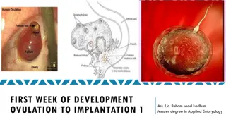
Development of the fertilized ovum Zygote Morula Blastocysts 6
Fetal development 0-4 weeks after conception o Rapid growth, formation of the embryonic plate o Primitive central nervous system forms o Heart develops and begins to beat o Covered with a layer of skin o Limb buds form 4-8 weeks o Very rapid cell division o Head and facial features develop o All major organs laid down in primitive form o External genitalia present but sex not distinguishable o Early movements visible on ultrasound from 6 weeks 6/17/2025 7
Fetal devt 8-12 weeks o Eye lids fuse o Kidneys begin to function and the fetus passes urine from 10 weeks o Fetal circulation functioning properly o Sucking and swallowing begin o Sex apparent o Moves freely (not felt by the mother) o Some primitive reflexes present 6/17/2025 8
Fetal devt... 12-16 weeks o Rapid skeletal development visible on x-ray o Meconium present in gut o Lanugo appears o Nasal septum and palate fuse 16-20 weeks o Quickening mother feels fetal movements o Fetal heart heard on auscultation o Fingernails can be seen o Skin cells begin to be renewed 6/17/2025 9
Fetal devt... 20-24 weeks o Most organs become capable of functioning o Periods of sleep and activity o Responds to sound o Skin red and wrinkled 24-28 weeks o Survival may expected if born o Eyelids reopen o Respiratory movements 6/17/2025 10
Feta devt... 28-32 weeks Begins to store fat and iron Testes descend in to scrotum Lanugo disappears from face Skin becomes paler and less wrinkled 32-36 weeks Increased fat makes the baby more rounded Lanugo disappears from body Head hair lengthens Nails reach tips of fingers Ear cartilage soft Plantar creases visible 6/17/2025 11
Fetal devt 37-40 weeks Term is reached and birth is due/expected Contour rounded Skull firm 6/17/2025 12
Summary of fetal development based on trimester First trimester (conception -12wk) Period of organogenesis Susceptible to teratogenic Heart functions at 3-4wks Eye formation at 4-5wks Arm and leg buds at 4-5wks Recognizable face and external genitalia at 8wk Placenta formed at 12wk Rapid growth of brain Bone ossified at 12wk 6/17/2025 13
Fetus at 8 Weeks 6/17/2025 14
Second trimester (13-24wk) Less susceptible for teratogens after 12wks Facial feature are formed at 16wk FHB heard by 16-20wks with fetoscope/Doppler Vernix present Size of fetus is around 25cm 6/17/2025 15
20 weeks (5 Months) 16 6/17/2025
Third trimester (25-36wks) Surfactant production begins in increasing amount Iron is stored Calcium stored at 28-32wks Fetal reflex at 28-32wks Subcutaneous fat deposits at 36wk Size of the fetus 45-55cm at 38-40wks 6/17/2025 17
Seven Months Eight Months 6/17/2025 18
Nine Months 6/17/2025 19
Summary of the fetal development based on gestational age A gestational age is the age of pregnancy counted in weeks. During the first 3 weeks of life the fertilized ovum is called a zygote. During this time cells have multiplied until Blastocyst has been formed and it has implanted itself in the walls of the uterus. During this time primitive trophoblastic villi have been formed to reach down into the thick endometrium thus giving contact with maternal blood. Starting from week 3 up to 8th week it is called Embryo. After 8th week up to delivery we call it a Fetus and after birth it is called a baby. 20
The Placenta The placenta is a round flat mass which is bluish red in color. It is about 20 cm in diameter and 2.5 cm in thickness from the center. It measures about 1/6 part of the baby s weight at birth. It links closely the fetus with the mother s circulation to carry out functions which the fetus is unable to perform itself during intrauterine life. 21
The placenta has two surfaces: - The maternal surface Is made up of chorionic villi arranged in 18 20 lobes or cotyledons. The maternal blood gives it a bluish red color The fetal surface The amnion covering the fetal surface of the placenta gives it a whitish, shiny appearance. Branches of umbilical vein and arteries are visible and spreading out from the insertion of the umbilical cord which is normally in the center. 22
There are two membranes on the placenta. The chorion: - It is the outer membrane of the placenta which is opaque, friable and adherent to the uterine wall until 3rd stage of labor. During the 3rd stage of labor, it detaches and expelled with the placenta. The amnion: - It is the smooth, tough and translucent inner membrane of the placenta. The fetus and the liquor are in it. It rupture during labor in order to expel the fetus. 23
Functions of the placenta 1.Nutrition: - The fetus gets the required nutrient from the mother through placenta. 2.Endocrine: - The placenta secrets different hormones by the cytotrophoblastic layer of the chorionic villi. A. Human Chorionic Gonadotrophic hormone (HCG) helps to maintain suitable condition within the uterus as the fetus develops. It also gives the basis for pregnancy test. 24
B/ Progesterone Its level rises from the 12th week and lasts throughout until the placenta is expelled It is used to prevent premature uterine contraction. C/ Estrogen Its production rises from the 6th week until term and fail after the expulsion of the placenta. It is used to enhance the mammary gland and enlargements of uterus. 25
D/ Human placental lactogens (HPL) Is a hormone w/c have a role in glucose metabolism in pregnancy It help to promote milk production and growth 3.Respiratory: - The fetal lung is not functional in the uterus so that the exchange of gas (CO2 & O2) is carried out by placenta. Oxygen from the mother s hemoglobin passes into fetal blood vessels by simple diffusion and similarly the fetus gives off CO2 into the maternal blood vessels. 4.Barrier (protection): - It provides a limited protection against infection. But few bacterial infections like syphilis and tubercle bacillus can penetrate. Some viruses, however, can cross freely and cause congenital abnormalities as in the case of the rubella and HIV virus. 26
5.Excretion: - The main substances excreted from the fetus is CO2 and bilirubin 6.Storage: - It metabolizes glucose, stores it in the form of glycogen & reconverts it back to glucose when needed. The placenta can also store iron & the fat soluble vitamin 27
The umbilical cord -Extends from the fetal umbilicus to the fetal surface of the placenta. -The umbilical cord encloses: - One umbilical vein - carrying oxygenated blood from the mother to the fetus Two umbilical arteries - carrying back deoxygenated blood from fetus to the placenta villi -Warthon`s jelly a sticky jelly like substance. -Its average length is about 50 cm and it is about 2 cm thick. 28
Types of umbilical cord insertion Central insertion: - is a normal type of the cord insertion w/c is in the middle of the placenta. Battledore insertion: - It is a type of cord insertion at the edge of placenta like tennis table bat. Velamentous insertion: - Here the cord is inserted in to the membranes some distance from the edge of the placenta. The umbilical vessels run through the membrane from the cord to the placenta. It is the dangerous type of cord insertion. Its major complication is detachment and tear of the blood vessels during active third stage of labor. As a result sudden blood lose will happen. 29
Placenta circumvallate: - An opaque ring is seen on the fetal surface; w/c means the chorion is attached on the fetal surface at some distance away from the edge. Placenta bipartite or tripartite: - When the placenta is divided in to two or three complete lobes each with a cord leaving it. The bipartite cord joins a short distance from the two parts of the placenta. As a result there may be retentions of the extra lobe during delivery. 31
Abnormal placenta Placenta succentureta:- A small extra lobe is present separated from the main placenta. It is joined to the main placenta by small blood vessels w/c run through the membranes to reach the placenta. It has a danger that this small lobe may be retained during delivery preventing uterine contraction resulting in PPH and infection 33
The amniotic fluid It is clear, pale straw colored liquor in w/c the fetus floats. The normal amount is from 500 ml 1500 ml It has 99% water, and 1% dissolved organic matters including: - -Food substances (protein, glucose, minerals, lipids), -Waste products (salt, urea, uric acid, creatinine) and -Hormones(insulin, prolactin,and rennin) It is slightly alkaline in PH (7.2) 34
Functions of the amniotic fluid Acts as a shock absorber to the fetus and prevents from injury Allow for the growth and free movement of the fetus It protects the fetus from change in temperature, because liquid changes temperature more slowly than air. It aids muscular development, because it allows the fetus freedom to move During labor it protects the placenta and umbilical cord from the pressure of uterine contraction Facilitate cervical effacement and dilation 35
Thank you!!! 36
Objective At the end of this session students are expected to : Recognize physiology of fetal circulation. Recognize the changes under goes after delivery Identify the regions on the fetal skull. Show important land marks on the fetal skull 38
The feotal circulation There is no mixture between the maternal and fetal blood. The fetus although in utero has its own circulatory system which is quite rudimentary i.e. immature, different from an adult circulation. Fetal blood has begun to exchange nutrients with the maternal circulation across the chorionic villi as early as 3rd weeks of intrauterine life. 39
During intrauterine life the fetal gastro-intestinal and respiratory systems are not functioning. So it obtains the necessary nutrient and oxygen from the mother through the placenta. There are several temporary structures in addition to the placenta itself and the umbilical cord. These enable the fetal circulation to take place but undergo changes after birth. 40
There are five temporary structures in the fetal circulation: The umbilical vein:-leads from the umbilical cord to the underside of the liver and carries blood rich in oxygen and nutrient to the fetus. The ductusvenosus: - vein to vein. -It connects the umbilical vein to the inferior vena cava. -At this point the oxygenated blood from the umbilical vein mixes with the deoxygenated blood returning back through the vena cava The foramen ovale: - an oval opening. -Temporary opening between the atria w/c allows the majority of blood entering from the inferior vena cava to the right atrium to pass directly in to the left atrium. 41
The ductusarteriosus: - artery to artery. - An opening w/c allows the mixture of the blood in the pulmonary artery to the blood in the aorta. -It is at a point where the pulmonary artery comes in to contact with the descending arch of the aorta. The hypogastric arteries: -These are the branches of the internal iliac artery. - Become the umbilical artery when they enter the umbilical cord. -They return the deoxygenated blood from the fetus to the placenta. 42
Physiologic changes at birth The main factor which helps the physiologic changes at birth is the establishment of respiration. On inspiration: - When the infant cries immediately after the birth, it means that the baby has taken the first breath. At this time the lungs expand and their field is increased. So the blood capillaries suck the blood in to the pulmonary circulation from the right ventricles. 45
The pulmonary veins now returns blood back to the left atrium and this causes the pressure in the left side of the heart to rise and become more than the pressure on the right side of the heart. This is responsible for the closure of the foramen ovale. The blood from the right ventricles finds its easiest way to the lung and so now flows directly to the lung. So this is responsible for the closure of the ductus arteriosus. 46
The cutting of the umbilical vein: - The clumping of the umbilical vein stops the flow of the blood in the umbilical vein, the hypogastric arteries, and this is responsible for the closure of the ductus venosus. This is also responsible for the reduction of pressure in the inferior vena cava and in the right side of the heart as well. 47
Changes on fetal circulation at birth 1. Ductus venous becomes the ligamentumvenosum 2. Foramen ovale becomes the fossa ovalis 3. Ductus arteries becomes the ligamentumarteriosum 4. Umbilical vein becomes the ligamentumteres 48
The Fetal Skull The Fetal skull is a bony box like cavity which contains and protects the delicate brain tissues. These delicate brain tissues may be injured during the birth process as they are subjected to great pressure. The feotal head is large in comparison to the true pelvis and it is the most difficult part to deliver whether it comes first or last. 49
For a feotal head to be delivered without trauma to the mother and the fetus, some adaptation between the fetal skull and maternal pelvis must take place during labor. Therefore; an understanding of the land marks and measurements of the fetal skull enables to recognize normal presentation and positions and facilitate delivery with least possible trauma to mother and child. 50


")
")
")


")


")