Alimentary system development involves the differentiation of structures from different germ layers. The alimentary system originates from endoderm, with the foregut, midgut, and hindgut forming distinct regions of the primitive gut tube. This process includes the formation of the mucus lining of the gut, musculature, fibrous stroma, and serous coat derived from various layers. Key structures like the mouth, anal canal, liver, stomach, and intestines develop from specific parts of the primitive gut tube. Blood supply is provided by major arteries such as the celiac artery, cranial mesenteric artery, and caudal mesenteric artery, ensuring proper vascular support during development.
Please find below an Image/Link to download the presentation.
The content on the website is provided AS IS for your information and personal use only. It may not be sold, licensed, or shared on other websites without obtaining consent from the author. If you encounter any issues during the download, it is possible that the publisher has removed the file from their server.
You are allowed to download the files provided on this website for personal or commercial use, subject to the condition that they are used lawfully. All files are the property of their respective owners.
The content on the website is provided AS IS for your information and personal use only. It may not be sold, licensed, or shared on other websites without obtaining consent from the author.
E N D
Presentation Transcript
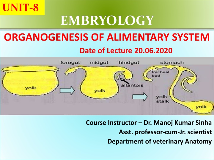
UNIT-8 EMBRYOLOGY ORGANOGENESIS OF ALIMENTARY SYSTEM Date of Lecture 20.06.2020 Course Instructor Dr. Manoj Kumar Sinha Asst. professor-cum-Jr. scientist Department of veterinary Anatomy
ALIMENTARY SYSTEM Develops from endoderm, endodermal layer forms mucus lining of gut and structure derived from gut Mouth, anal canal are derived from ectoderm The musculature, fibrous stroma and serous coat derived from the splanchnopleuric layer of mesoderm Primitive gut tube extends along the mid line from cephalic end (bucco-phararyngeal) to the caudal end (cloacal membrane) The primitive gut tube is derived from the dorsal part of the yolk sac Primitive gut tube is divided into three section : Fore gut, Mid gut and Hind gut
FORE GUT It is cranial part of the gut tube and separated from the stomatodaeum (primitive mouth cavity) from buccopharyngeal membrane at early stage Later at 4th week of development this bilaminar membrane disappears Caudally fore gut extends upto 2nd part of duodenum at the level of openinig of bile duct Fore gut divided into two parts : pre-laryngeal Part and post- laryngeal parts give rise to following parts: Pre-pharyngeal part Post-pharyngeal part Part of mouth cavity, Esophagus, stomach ,liver Pharynx, tongue, glands, plaatine pancreas, proximal part of tonsils, respiratory passage duodenum
MID GUT This is middle part of primitive gut tube which extends from duodenum to transverse colon in adults Mid gut connected with the extraembryonic part of the yolk sac by vitello-intestinal tube Mid gut divided into two parts: pre-cecal part and post-cecal part Pre-cecal Distal part of duodenum cecum, colon Jejunam, Ileum (ascending and transverse) Post-cecal
HIND GUT It is the caudal part of the primitive gut tube, and separated from the ectoderm by clocal membrane (bilaminar membrane) Hind gut is divided into two parts: pre-allantoic part and post allantoic part pre-allantoic part post allantoic part Descending colon Primitive urogenital sinus Rectum The splanchnopleuric layer of the mesoderm after enclosing the gut with its muscular, fibrous and serous layer forms primitive dorsal mesentery The dorsal mesentery suspends the gut from the dorsal wall and gives passage to blood vessels Celiac artery supplies the abdominal part of the fore gut, cranial mesenteric artery supplies the mid gut and caudal mysentric artery supplies the hind gut
PRE-LARYNGEAL PART OF THE FORE GUT Cephalic part of fore gut separated from stomatodaeum by buccopharyngeal membrane and after rupture of this membrane cephalic part of foregut opens to the exterior through stomadodaeum Then primitive pharynx is formed and is composed of three layer: Ectoderm, Mesoderm and Endoderm Mesoderm of the lateral wall of fore gut undergoes thickening at intervals and form a series of bars (6 pairs) k/a Branchial or Pharyngeal arches , each of these bars grow and take the shape of arch k/a Pharyngeal Arches The grooves which develop outside between the bars are k/a Branchial clefts and those develop outside between the bars are k/a Pharyngeal pouches Branchial arches + Branchial clefts + Pharyngeal pouches together called Branchial Apparatus Branchial Apparatus resposible for development of face, neck, mouth, pharynx and larynx
BRANCHIAL CLEFTS AND PHARYNGEAL POUCH BRANCHIAL CLEFTS First cleft gives rise to the lining membrane of External acoustic meatus 1st 2nd and 4th clefts invaginate into mesenchyme and form placodal vesicles These vesicles associated with the development of ganglia of 7th 9th and 10th cranial nerves PHARYNGEAL POUCH: 1st pouch and part of 2nd Forms Auditary tube and tympanic cavity 2nd pouch (part) Forms Tonsil 3rd pouch Forms Inferior Parathyroid and Thymus 4th pouch Forms superior parathyroid and thyroid gland 5th pouch Forms Uitimobronchial body that gives rise to parafollicular part of the thyroid gland
BRANCHIAL ARCHES Derivatives of Branchial Arches ARCHES ARTERIES NERVES SKELETON MUSCLES 1st (Mandibular Arch) Part of Maxillary artery Mandibular, chorda tympani Maxillary process, mandible, malleus, incus Muscles mastication, Tensor tympani of 2nd (Hyoid Arch) - Facial Body of hyoid bone,Stapes Stapedius, stylohyoid and facial muscles 2rd Arch Commomn carotid Glossopharryn geal Great cornu of hyoid arch Stylophryngious 3rd Arch Arch of Aorta Superior Laryngeal Thyoid cartilage Cricothyoid 4th Arch Disappear early 5th Arch Pulmonary artery, arteriosus Recurrent laryngeal Cricoid arytenoid cartilage and Intrinsic muscles of larynx execpt Crico-thyroid Ductus