Embryonic Membranes and Development in Veterinary Anatomy
Explore the development of extraembryonic membranes like the amnion, yolk sac, and allantois in veterinary embryology. Understand their role in protecting and nourishing the embryo throughout different animal species. Dive into the intricate processes of placenta formation and embryonic growth under the instruction of Dr. Manoj Kumar Sinha.

Download Presentation

Please find below an Image/Link to download the presentation.
The content on the website is provided AS IS for your information and personal use only. It may not be sold, licensed, or shared on other websites without obtaining consent from the author. If you encounter any issues during the download, it is possible that the publisher has removed the file from their server.
You are allowed to download the files provided on this website for personal or commercial use, subject to the condition that they are used lawfully. All files are the property of their respective owners.
The content on the website is provided AS IS for your information and personal use only. It may not be sold, licensed, or shared on other websites without obtaining consent from the author.
E N D
Presentation Transcript
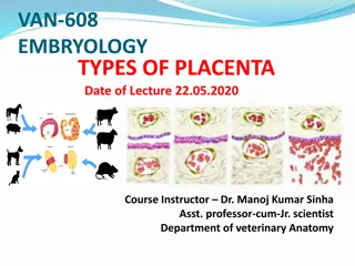
Course No-VAN608 (MVSC) Topic - General embryology (Development of placenta) Course instructor Dr. Manoj Kumar Sinha Department of veterinary Anatomy
EXTRAEMBRYONIC MEMBRANE During development of embryo the germ layer extends far from the embryonic area and peripheral part of the germ layer give rise to extra embryonic membrane It is discarded at the time of birth Four types of extra embryonic membrane: Amnion II. Yolk sac III. Allantois IV. Chorion I.
AMNION Develops into folds from all parts of embryo Due to continuous growth these folds came together above the mid dorsal region of embryo and fuse with one another to form a single covering of somatopleure The limb of somatopleure forms amnion and outer limb forms chorion (serosa) Tracheal and oral secretion as well as urine enters into amniotic sac as pregnancy advances The embryo remain suspended in amniotic fluid which prevent its adhesions and prevent the embryo from injury The amniotic sac is fully formed in pig, sheep and cow in 16th17thand 18thdays of fertilization respectively In chick it is formed in 72 hrs of incubation
YOLK SAC It is the first extra embryonic membranes appear There are three folds : cephalic, lateral, and caudal fold these folds push the somatopleure downward and affect the splanchnoplerue Anterior part of endoderm forms-Foregut Middle undifferentiated part forms-Hind gut Original endodermal sac continuous with midgut is called yolk sac In cattle and pig it absorbed the uterine milk to provide nutrition to development embryo. In horse & dog it develops villi. In adult remnant of yolk sac present at the junction of ileum & jejunum as small diverticulum diverticulum . It is more common in birds and ruminants. remains undifferentiated and called Meckel s
ALLANTOIS It develops from the splanchopleure . It appear as a diverticulum from ventral wall of hindgut on its external surface. Its narrow proximal part is called allantoic stalk & distal portion is called allantoic vesicle. In dog & horse its completely encloses the embryo. In pig and sheep it does not cover the dorsal aspect of embryo. Allantoic fluid smooth discoidal rubber like brown masses make their appearance during later part of pregnancy is called Hippomanes. In pig allantois appear on 6thday & fully formed by 28 day of post fertilization & chick appear on 3rdday & full formed 9th day. It dispose of waste products & contributes to gas exchange.
CHORION Chorion is outermost extra embryonic membrane. It is a composite structure formed due to fusion of allantois & serosa. It consist of four layers. a. Trophoectoderm b. Somatic mesoderm c. Splanchnic mesoderm d. Endoderm The allantois fuses with serosa in cow, sheep, and pig on 36th day In the mares a girdle like structure is formed in chorion, cells of this girdle migrated into endometrium and transformed into concave shaped structre called Endometrial cups Endomerial cups are 12 in number and it secrets equine chorionic gonadotropin harmone kown as PMSG (Pregnant Mare Serum Gonadotropin)
")