Enteric Fever (Typhoid Fever)
Enteric fever, caused by Salmonella typhi, and bacillary dysentery, caused by Shigella, are two gastrointestinal infections with distinct etiologies, clinical manifestations, pathogenesis, and treatment options. Understanding these diseases is essential for effective management and prevention strategies. Explore the key differences between these infections, their laboratory diagnosis, and treatment approaches.

Download Presentation

Please find below an Image/Link to download the presentation.
The content on the website is provided AS IS for your information and personal use only. It may not be sold, licensed, or shared on other websites without obtaining consent from the author.If you encounter any issues during the download, it is possible that the publisher has removed the file from their server.
You are allowed to download the files provided on this website for personal or commercial use, subject to the condition that they are used lawfully. All files are the property of their respective owners.
The content on the website is provided AS IS for your information and personal use only. It may not be sold, licensed, or shared on other websites without obtaining consent from the author.
E N D
Presentation Transcript
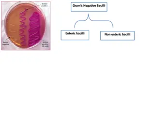
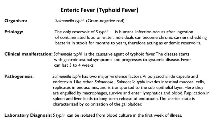
Enteric Fever (Typhoid Fever) Organism: Salmonella typhi (Gram-negative rod). Etiology: The only reservoir of S typhi of contaminated food or water.Individuals can become chronic carriers,shedding bacteria in stools for months to years, therefore acting as endemic reservoirs. is humans.Infection occurs after ingestion Clinical manifestation: Salmonella typhi is the causative agent of typhoid fever. The disease starts with gastrointestinal symptoms and progresses to systemic disease. Fever can last 3 to 4 weeks. Pathogenesis: Salmonella typhi has two major virulence factors, Vi polysaccharide capsule and endotoxin. Like other Salmonella , Salmonella typhi invades intestinal mucosal cells, replicates in endosomes, and is transported to the sub-epithelial layer. Here they are engulfed by macrophages, survive and enter lymphatics and blood. Replication in spleen and liver leads to long-term release of endotoxin. The carrier state is characterized by colonization of the gallbladder. Laboratory Diagnosis:S typhi can be isolated from blood culture in the first week of illness.
Treatment and Prevention: a variety of antibiotics can be used to control the course of infection including: Ampicillin, Chloramphenicol, Trimethoprim, Ceftriaxone, and Ciprofloxacin. Prevention involves proper sanitation, carriers not handling food and vaccination against Vi polysaccharide.
Bacillary Dysentery Organism: Shigella (Gram-negative rod ,4 species: S dysentericae, S flexneri, S bodydii, and S sonnei). Etiology: Transmission is most often by feca-oral spread.Very few organisms (100-200) are required to cause infection. Clinical manifestation: Shigella causes dysentery that is clinically similar to that caused by enteroinvasive E coli (EIEC). Symptoms include fever, abdominal cramps, and blood and mucous diarrheal stool . S dysentericae also produces Shiga toxins, which is associated with more serious disease and development of hemolytic uremic syndrome. Pathogenesis: : Shigella carries a number of virulence factors including genes required for invasion, endotoxin and Shiga toxin. As with Salmonella ,Shigella invades through intestinal mucosal cells. It then escapes the endosomes, replicates in the cytoplasm, spreads to the adjacent enterocytes. Cell destruction induces a host inflammatory response.Shiga toxin is not required for dysentery. Laboratory Diagnosis: like Salmonella, Shigella are lactose fermenters and can be isolated on a variety of selective and differential media from stool cultures. Shigella do not produce H2S and are nonmotile. Treatment and Prevention: Fluid and electrolyte replacement is often adequate for mild cases, whereas antibiotics such as ciprofloxacin and trimethoprim can be used for more serious disease. Prevention involves proper sanitation and good personal hygiene.
Treatment and Prevention: Fluid and electrolyte replacement is often adequate for mild cases, whereas antibiotics such as ciprofloxacin and trimethoprim can be used for more serious disease. Prevention involves proper sanitation and good personal hygiene.
Organism: Pseudomonas aeruginosa (Gram-negative rod).Strict aerobes, can grow in distilled water with few nutrients. Etiology and epidemiology: This ubiquitous organism can be found in soil,water,plants ,and food.It is a common contaminant on numerous objects in hospital and can survive in many disinfectants. Clinical manifestation: Pseudomonas infections are opportunistic with major manifestations including wound infections in burn patients, recurring pneumonia in cystic fibrosis patients, and urinary tract infections associated with indwelling catheters. Pathogenesis: : Virulence factors include endotoxin, exotoxin A, exoenzyme S, adhesions, and antiphagocytic capsule. Conditions that facilitate colonization are important in pathogenesis.Examples include mucus production in cystic fibrosis that inhibits ciliated clearance and burns that remove skin barriers. ExotoxinA is an A-B toxin with the same mechanism of synthesis inhibition and cell death as diphtheria toxin. Exoenzyme S is an ADP-ribosylatingprotein toxin that targets several cellular proteins. Laboratory Diagnosis: When grown on normal laboratory media, Pseudomonas gives off a fruity aroma. It also produces several pigments: pyocyanin gives a blue color to pus: pyoverdin is a yellow- green molecule that is fluorescent under ultraviolet light and can be used to monitor burn patients for infection.
Treatment and Prevention: Sensitivity testing is important because Pseudomonas is resistant to many different antibiotics. Prevention involves physical isolation and sterile practices of susceptible individuals.
")