Learn about Enterobious Vermicularis, also known as pinworm or threadworms, including its distribution, transmission, life cycle, and common infections in children. Discover how this parasite is transmitted, its symptoms, and preventive measures to protect against infection.
Please find below an Image/Link to download the presentation.
The content on the website is provided AS IS for your information and personal use only. It may not be sold, licensed, or shared on other websites without obtaining consent from the author. If you encounter any issues during the download, it is possible that the publisher has removed the file from their server.
You are allowed to download the files provided on this website for personal or commercial use, subject to the condition that they are used lawfully. All files are the property of their respective owners.
The content on the website is provided AS IS for your information and personal use only. It may not be sold, licensed, or shared on other websites without obtaining consent from the author.
It has world wide distribution. It is common in cold climate than worm climates, because of less frequent bathing & infrequent changing of underwear cloths. It is the most common infection in united states.
Transmission E.Vermicularis transmitted by ingestion of infective eggs. The eggs are deposited on anal skin usually during night & within few hours of being laid they contain infective larva. ` Infection is easily spread by clothing & bedding. Autoinfection is common in children because eggs cause intense irritation of the infected area lead to contamination of fingers
Occasionally larvae hatch from the eggs on the perianal skin and migrate back into the intestine where they grow into mature worms. This type of infection is called retro infection. Airborne transmission of E. vermicularis can also occur.
Life cycle The life cycle start when the human ingest the infective eggs. The eggs hatch in small intestine & develop to mature worms in large intestine. After mating ,the female migrate to rectum, pass out of the anus & lay their eggs on the perianal skin. Within 6 hours each egg contain an infective larva. The eggs produce 6 week after infection. Depending on climatic condition ,the infective eggs can remain viable on clothing for several weeks .
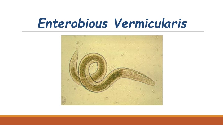
Adult worms There are small, yellow- white . The female measures 8 13 mm in length & has thin pointed tail . The male measure 2 5 mm & has coiled tail with single spicule.
Clinical feature & pathology E.Vemicularis rarely causes serious symptoms ,usually there is intense irritation round the anus. In female ,infection of urinary & genital tract may occur. Worms in appendix may associated with appendicitis.
Diagnosis Confirmation of enterobiasis by : Finding eggs in perianal skin samples, faeces & less commonly in female urine . Finding of worms in faeces.
Clear adhesive tape technique This method used to collect sample from perianal skin. High number of eggs can be usually recovered in the morning after waking & before bathing. After collecting the eggs, stick the tape lengthways, face down on a microscope slide. Immediately before examining the preparation, lift up the tape and add a drop of xylene to the center of the preparation. Press down the tape again, smoothing it out with a piece of cotton.
Use of xylene: This is not essential but a drop of xylene will clear any debris and air bubbles, making it easier to detect the eggs. Do not, however, add the xylene until ready to examine the preparation because prolonged contact with xylene may cause the eggs to collapse. Adhesive tape-slide preparations of E. vermicularis keep well for several days when stored at 2 8 C in case the examination of the specimen need to be delayed.
4. Examine the preparation microscopically using the 10 objective to detect the eggs and the 40 objective to identify them. The condenser iris must be closed sufficiently to give good contrast otherwise the colourless eggs will not be seen.
Caution Most preparations will contain infective eggs by the time they reach the laboratory for examination. Pinworm eggs are frequently plentiful and can be carried by air currents. Each egg will often contain a fully developed embryo and will be infective within a few hours after being deposited. Wear gloves and consider the use of a face mask if you suspect the sample will allow eggs to become airborne.
Swab method 1. Moisten a cotton wool swab in fresh physiological saline. 2. Swab around the perianal area. 3. Agitate the swab in a container of 4 5 ml of saline to wash off the eggs. 4. Cap the container and safely discard the swab. 3 Transfer the saline to a conical tube and centrifuge to sediment the eggs. 4. Discard the supernatant fluid and without delay, transfer the sediment to a slide, cover with a cover glass and examine microscopically.
Adhesive tape preparation showing eggs ofE.vermicularis recovered from anal skin.
Egg of E.vermicularis It is colourless & has clear shell. Oval & usually flattened on one side (D shape) . Measure about 50 x30 m .
Prevention & control Because E.vermicularis eggs are infective very soon after being laid, the family or community often becomes infected ,therefore prevention & control include : Treating all family members . Washing of anal skin each morning soon after waking. Washing of cloths worn at night separately & by worm water.