Epidemiological Study of Setaria equina Infection in Donkeys
Setaria equina is a common vector-borne pathogen in equines worldwide, with potential effects on donkeys including peritonitis and neurological disturbances. This study aimed to investigate the prevalence of S. equina in central Iraq and identify the worm through morphological features under a light microscope.

Download Presentation

Please find below an Image/Link to download the presentation.
The content on the website is provided AS IS for your information and personal use only. It may not be sold, licensed, or shared on other websites without obtaining consent from the author.If you encounter any issues during the download, it is possible that the publisher has removed the file from their server.
You are allowed to download the files provided on this website for personal or commercial use, subject to the condition that they are used lawfully. All files are the property of their respective owners.
The content on the website is provided AS IS for your information and personal use only. It may not be sold, licensed, or shared on other websites without obtaining consent from the author.
E N D
Presentation Transcript
Epidemiological study of Setaria equina infection in donkeys . . . .
Introduction Setaria equina (S. equina) is a common vector borne pathogen of equines all over the world, especially in tropical zones. S. equina transmitted by Aedes aegypti and Culex pipens where L1 developed to L3 within 2 weeks in their thoracic muscles. The equines acquired the infection during mosquito s blood meal and the life cycle completed within 8- 10months [Perumal et al., 2016]. Prenatal infection reported as a route of transmission for Setaria species [Kim et al., 2010; Marzok & Desouky , 2009]. Adult worms mainly found in the peritoneal cavity of horse and donkey. The worms were nonpathogenic but might induce varied degrees of peritonitis and might migrate to the eye, brain, lung, and scrotum of equines causing lacrimation, blindness, paraplegia, locomotor, and neurological disturbances [Solusby, 1986 ; Rhee et al ., 1994]. S. equina induced such pathogenic effects but also other Setaria species infect cattle (S. digitata and S.cervi) could induce blindness and CNS damage in equine where [Shin et al ., 2017]. The parasite reported the aberrant parasitism of adult worms in eye of donkey in Egypt. [Abu El-Magd, & AHMED, 1994] Also, its stated the zoonotic importance of S. equina in man.[Taylor,2001; Abbas et al., 2016]
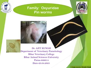
Morphology Male measurements reached 45-70mm (57 2) long 0.4- 0.6 mm (0.45 0.02) wide with coiled end and Female reached 60-160mm (110 5) long 0.6-0.91mm (0.60 0.04) wide in females with loose spiral end. [Abdel Rahman & M.M.I. 2020]. The larval stage of Microfilaria in Knott s technique examination Fig. 1,2 Fig. 1 Male (m) and female (f) worms. Fig. 2 Microfilaria in Knott s technique ( 100).
Aims of the study The present study aimed to assess the following : Investigate the prevalence rate of S. equina in donkeys in central part of Iraq. Identify the worm by morphological features of the genus Setaria under the light microscope
Methodology Methodology Animals A total of 218 (106 male and 112 female) animals slaughtered in Baghdad ( Al-Zawra ) Zoo, brought from of central part of Iraq (Diyala, Wasit, Baghdad). The Animals were examined during the period from December 2012 to August 2010. Sample collection and processing Adult worms were collected from peritoneal cavity of donkeys at the time of necropsy, washed with saline, cleared with lactophenol, and identified under light microscope according to [Burgu et al., 1995]. The data was analyzed by using the statistical program SAS .
Results The total infection rate of Setaria equina was 11.9% and the highest infection rate (22.2%) was recorded in October, where is no infection in January, February and August. The high infection rate (14.5% ) during the months of spring and autumn (18.8%) and decrease in Winter and Summer 3.6% and 10%. Tab. 1 Tab.1
40 X Results Adult worms inside the peritoneum of donkeys appeared milky white and thread like. The worms was distinguished and confirmed the diagnosis by observing the distinctive morphological Horn-like Structure characteristics of the worm during microscopic examination and the shape of the front end of males and females. Fig. 3 Fig.3 The anterior end
x40 Results Fig.4 Male posterior end Male and Female posterior end shows the x 40 morphological characteristics of the worm by microscopic examination . Fig.4,5 Fig 5. female posterior end
Results Female donkeys with a higher proportion (13.5%) than male10.3% respectively differences ( P < 0.05 ). Fig. 6 Worm intensity ranged beween 1-6 worm and was shown high worm burden in Autumn and Spring fallowed by Summer and Tab.2 Finally Winter seasons ,which no infection was recorded in some months Tab.2 Fig. 6
These worms were first recorded in Iraq by Leiper, (1957) and limited numbers of examined horses at thet time. Ali et al., 1992; has recorded by 10.5% when conducting the autopsy of 124. Al-Al-Alusy et al., 1994. found these worms in fifty of the horses of the city of Mosul through blood tests were able to detecting the worm diagnosed larval phases (nematodes). While, other fallowing didn t recorded any similar results for the difficulty of conducting the autopsy and investigation of adult worms) Al-Falahay , et al., 1992. Discussion Many species of Setaria were reported all over the world, but S.equina considered the most popular species recorded in donkeys. In Iraq, S.equina had not been studied by many authors and this study were cited by 11 world and middle east studies and mainly in Egypt.
Conclusion The worms of the genus Seteria represent economic importance. The diseases may cause [Cerebrospinal Nematodiasis], and this is no less important in the equine in Iraq and the rest of the world. Recommendations 1. Using scanning electron microscopy considered a good choice to distinguish the ultrastructures . 2. Perform the phylogenetic analysis and detect relationships between different filarial worms, which could not detect by the morphological characterization of adult worms. 3. The need of the programs for proper and an effective control strategy against filariasis specially S. equina in Iraq. 4. The diagnosis of S. equina microfilariae in blood samples by PCR will be crucial for rapid and effective treatments .