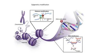
Epigenetics of Atherosclerosis

The research delves into epigenetic influences on atherosclerosis using longitudinal studies and repeat data collections on monocytes and carotid plaques. Investigating genomic features in predicting plaque vulnerability and CVD risk factors.

Download Presentation

Please find below an Image/Link to download the presentation.
The content on the website is provided AS IS for your information and personal use only. It may not be sold, licensed, or shared on other websites without obtaining consent from the author.If you encounter any issues during the download, it is possible that the publisher has removed the file from their server.
You are allowed to download the files provided on this website for personal or commercial use, subject to the condition that they are used lawfully. All files are the property of their respective owners.
The content on the website is provided AS IS for your information and personal use only. It may not be sold, licensed, or shared on other websites without obtaining consent from the author.
E N D
Presentation Transcript
Epigenetics of Atherosclerosis Yongmgei Liu, MD, PhD, FAHA Wake Forest School of Medicine Claudia E. Korcarz, DVM Atherosclerosis Imaging Research Program University Wisconsin
MESA Epigenomics Study in Exam 5 MESA epigenomics study funded by NIH Roadmap Epigenomic Program and NHLBI ~2500 participants WFU, JHU, Columbia, UMN Identify genomic features (DNA methylation and resultant mRNA expression) in monocytes (involved in atherogenesis) as novel risk factors or biomarkers of atherosclerosis Limitation of the cross-sectional study design Warrants a prospective follow-up study
A longitudinal Epigenetics Study of Atherosclerosis MESA Exam 6 (R01 HL135009-01 ) Aim 1. To test predictive effects of candidate genomic features on initiation/progression of atherosclerosis New carotid plaque Change in carotid plaque scores between the two exams Aim 2. To characterize the associations of the promising genomic features with plaque vulnerability Carotid plaque echolucency and other characteristics Aim 3. To identify temporal and causal relationships between CVD risk factors (genetic or traditional), the promising genomic features, and plaque burden
Sample/Data Collection Repeat epigenomic and transcriptomic profiling of monocytes in Exam 6 ~2500 participants WFU, JHU, Columbia, UMN CBC/diff, PBMC/monocyte/T cell purification DNA methylomic profiling using sequencing technology Transcriptomics: mRNA-seq and total RNA-seq Repeat measures of known CVD risk factors Fasting glucose, HbA1C, insulin, creatine etc.
Sample/Data Collection Repeat carotid ultrasonography in Exam 6 All returning participants who had ultrasonography at Exam 5 (N=~1,500) WFU, JHU, Columbia, UMN B-mode ultrasound images Read ultrasound images at UW AIRP Director: James Stein, MD Manager: Claudia E. Korcarz, DVM
Exam 6 Ultrasound Equipment and Protocol 4 new Mindray M9s- GE Logiq 700 not serviceable Abilities to detect plaque and derive plaque scores (binary, ordinal measures) do not differ qualitatively or quantitatively from E5 Nearly identical transducer frequency Carotid plaque = discrete lesion 1.5 mm or focal thickening 50% Wall thicknesses are >0.7 mm, plaques tend to be at least 1 mm thick which is much greater than the resolution of previous and current ultrasound systems Verified on small parts phantom & DICOM image file data
Study Updates US systems shipped and installed (2-3/2017) All FC completed trainings Web-based lectures 2 days on-site hands-on training 8 sonographers certified Major hurdle dealing with FC-specific security issues regarding image transfer and speed of transfer all troubles resolved As of April 6, 2017 we have received N=114 studies
Carotid Scans Performed by 4/6/2017 Potential Participants with Exam 5 data Field Center Mean (SD) quality score Failed exams n,(%) # Scans COL 549 5 96.0 (5.5) 0 (0%) JH 368 48 98.3 (4.8) 0 (0%) UMN 500 22 84.1 (17.3) 3 (13%)** WFU 475 39 88.2 (9.7) 2 (5.1%) TOTAL 1,892 114 92.0 (11.5) 5 (4.4%) **Providing additional on-site training and help with scanning participants for one sonographer
Plaque Detection and Plaque Score Reproducibility for carotid plaque detection and plaque score in E5 was excellent Intra-reader ( =0.83, 95% CI 0.70 0.96) Inter-reader ( =0.89, 95% CI 0.72 1.00) The 2 readers assigned for E6 were readers for E5 E5 images reviewed in cases of discrepant findings QC for inter- and intra-scorer reproducibility will be tested in an ongoing basis until the end of Exam 6 Tattersall MC et al. Stroke 2014;45(11):3257-62 Gepner AD et al. Circ Cardiovasc Imaging 2015;8(1)
Exam 5 vs. Exam 6 Left Bulb far wall plaque
Carotid Plaque Echogenicity Analysis Images normalized and standardized to a pixel density of 20/mm Image crop and feature extraction modules to measure GSM, plaque area Just black areas manually traced; area calculated Discrete white areas are noted Black areas adjacent to the lumen with an echogenic border are noted Drs. Mitchell and Dempsey validated these measures against autopsy specimens Mitchell CC et. al. Ultrasound in Med Biol 2017; 43(1):129
High GSM 184, Plaque area 5.95 mm2 Low GSM 19, Plaque area 50.77 mm2
Ultrasound Plaque Characteristics Measurement Reproducibility - MESA Exam 1 plaques Reader 1 Reader 2 Reader 1 vs Reader 2 Intra- observer Mean Delta Intra- observer Mean Delta Inter- reader mean Delta Within Subjects SD Within Subjects SD Within Subjects SD ICC ICC ICC Variable 0.96 (0.92- 0.98) 0.99 (0.98- 1.00) 0.96 (0.92- 0.98) 0.95 (0.88- 0.98) 0.98 (0.95- 0.99) 0.81 (0.61- 0.91) 0.95 (0.88- 0.98) 0.98 (0.95- 0.99) 0.86 (0.70- 0.94) GSM (units) 0.96 4.39 5.22 5.30 5.7 5.53 Plaque Area (mm2) Black Areas (mm2) 0.20 1.11 1.82 1.67 1.93 1.66 0.13 0.37 1.54 0.77 0.37 0.61 24 plaque images from MESA Exam 1 were re-measured by the same 2 readers assigned for Exam 6 data analysis
QC Plan for Exam 6 Data The DCC will select a set of 20 studies/FC (n=80) to be re-scored for plaque presence and plaque score. The representative set will be selected after each FC has scanned their first 50 MESA participants to assess intra- and inter-reader reproducibility The DCC will randomly select a representative set of 60 plaques (15/FC) to assess intra- and inter-reader reproducibility for the plaque characteristic measures Reader reproducibility will be repeated every 8 months until the end of Exam 6 reading phase