Epithelial Cell Adhesion & Junctions in Tissues
Epithelial cells exhibit strong intercellular adhesion through specialized junctions, serving as barriers, adhesion sites, and communication channels. These cells form sheets that cover surfaces, absorb nutrients, secrete substances, and more. Their unique features and functions make them crucial for bodily functions.

Download Presentation

Please find below an Image/Link to download the presentation.
The content on the website is provided AS IS for your information and personal use only. It may not be sold, licensed, or shared on other websites without obtaining consent from the author.If you encounter any issues during the download, it is possible that the publisher has removed the file from their server.
You are allowed to download the files provided on this website for personal or commercial use, subject to the condition that they are used lawfully. All files are the property of their respective owners.
The content on the website is provided AS IS for your information and personal use only. It may not be sold, licensed, or shared on other websites without obtaining consent from the author.
E N D
Presentation Transcript
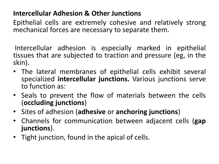
Intercellular Adhesion & Other Junctions Epithelial cells are extremely cohesive and relatively strong mechanical forces are necessary to separate them. Intercellular adhesion is especially marked in epithelial tissues that are subjected to traction and pressure (eg, in the skin). The lateral membranes of epithelial cells exhibit several specialized intercellular junctions. Various junctions serve to function as: Seals to prevent the flow of materials between the cells (occluding junctions) Sites of adhesion (adhesive or anchoring junctions) Channels for communication between adjacent cells (gap junctions). Tight junction, found in the apical of cells.
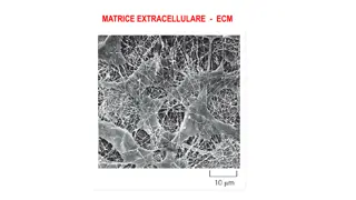
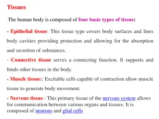
Epithelial tissues Epithelial tissues are composed of closely aggregated polyhedral cells with very little extracellular substance. These cells have strong adhesion and form cellular sheets that cover the surface of the body and line its cavities. The principal functions of epithelial tissues are: Covering, lining, and protecting surfaces (eg, skin) Absorption (eg, the intestines) Secretion (eg, the epithelial cells of glands) Contractility (eg, myoepithelial cells). Specific cells of certain epithelia are also highly specialized sensory cells, such as those of taste buds or the olfactory epithelium. Because epithelial cells line all external and internal surfaces of the body, everything that enters or leaves the body must cross an epithelial sheet.
Characteristic Features of Epithelial Cells The forms and dimensions of epithelial cells range from high columnar to cuboidal to low squamous cells. Epithelial cell nuclei have a distinctive shape, varying from spherical to elongate. The nuclear form often corresponds roughly to the cell shape; thus, cuboidal cells have spherical nuclei, and squamous cells have flattened nuclei. The long axis of the nucleus is always parallel to the main axis of the cell. Most epithelia rest on connective tissue. In the case of epithelia lining the cavity of internal organs (especially in the digestive, respiratory, and urinary systems) this layer of connective tissue is often called the lamina propria. The lamina propria not only serves to support the epithelium but also provides nutrition and binds it to underlying structures.
Specializations of the Apical cell Surface: Microvilli In absorptive cells, such as the lining epithelium of the small intestine, the apical surface presents orderly arrays of many hundreds of more permanent microvilli , the total surface area can be increased as much as 20- or 30-fold. The complex of microvilli and glycocalyx is easily seen in the light microscope and is called the brush or striated border. Stereocilia are long apical processes of cells in other absorptive epithelia such as that lining the epididymis and ductus deferens. These structures are much longer and less motile than microvilli, are branched, and should not be confused with true cilia. Like microvilli, stereocilia also increase the cells' surface area, facilitating the movement of molecules into and out of the cell.
Cilia Cilia are elongated, highly motile structures on the surface of some epithelial cells, which is much longer and two times wider than a typical microvillus. organisms, cilia exhibit rapid back-and-forth movements coordinated to propel a current of fluid and suspended matter in one direction over the ciliated epithelium. A ciliated cell of the trachea lining is estimated to have about 250 cilia. In living Flagella Present in the human body only in spermatozoa, are similar in structure to cilia but are much longer and are normally limited to one flagellum per cell.
Simple squamous epithelia. In simple squamous epithelium, cells of the single layer are flat and usually very thin, with only the thicker cell nucleus appearing as a bulge to denote the cell. Simple epithelia are typically specialized as lining of vessels and substances which can enter underlying tissue from the vessel or cavity. cavities and regulate Simple cuboidal epithelium. Cells of simple cuboidal epithelia vary in their height but are roughly as tall as they are wide. Examples a renal collecting tubule, a pancreatic duct, and the mesothelium covering an ovary
Simple columnar epithelium. Cells of simple columnar epithelia are taller than they are wide. Such cells are usually highly specialized for absorption, with microvilli, and often have interspersed secretory cells or ciliated cells. The examples shown here are from a renal collecting duct, the oviduct lining, with both secretory and ciliated cells, and the lining of the gall bladder.
Stratified squamous The very thin surface cells of stratified squamous epithelia can be "keratinized" (rich in keratin intermediate filaments) or "nonkeratinized. Stratified squamous keratinized epithelium is found mainly in the epidermis of skin. Its cells form many layers, and the cells closer to the underlying connective tissue are usually cuboidal or low columnar. The cells become irregular in shape and flatten as they accumulate keratin in the process of keratinization and are moved progressively closer to the surface, where they become thin, metabolically inactive packets (squames) of keratin lacking nuclei. This surface layer of cells helps protect against water loss across this epithelium. Stratified epithelium lines wet cavities (eg, mouth, esophagus, and vagina). In such areas where water loss is not a problem, the flattened cells of the epithelial surface layer are living cells containing much less keratin and retaining their nuclei. squamous nonkeratinized
Stratified cuboidal and stratified columnar epithelia are rare. Stratified columnar epithelium can be found in the conjunctiva lining the eyelids, where it is both protective and mucus secreting. Stratified cuboidal epithelium is restricted to large excretory ducts of sweat and salivary glands, where it apparently provides a lining more robust than that of a simple epithelium. Transitional epithelium or urothelium, which lines only the urinary bladder, the ureter, and the upper part of the urethra, is characterized by a superficial layer of domelike cells that are neither squamous nor columnar. These cells, sometimes called umbrella cells, are essentially protective against the hypertonic and potentially cytotoxic effects of urine. Importantly, the form of the surface cells changes according to the degree of distention of the bladder wall.
pseudostratified columnar epithelium, so called because all cells are attached to the basal lamina even though their nuclei lie at different levels in the epithelium and the height of some cells does not extend to the surface. The best- known example of pseudostratified columnar epithelium is that lining the passages of the upper respiratory tract. The columnar cells of this epithelium are also heavily ciliated.
Figures:1.simple squamous 2. Simple cuboidal 3. Simple columnar (ciliated, non ciliated) 4. Stratified squamous non keratinized 5. Pseudo stratified 6. Transitional epithelial
Glandular Epithelia Glandular epithelia are formed by cells specialized to secrete. The molecules to be secreted are generally stored in the cells in small membrane-bound vesicles called secretory granules. Glandular epithelial cells may synthesize, store, and secrete: Proteins in the pancreas Lipids in adrenal, sebaceous glands or Complexes of carbohydrates and proteins salivary glands. Mammary glands secrete all three substances. The cells of some glands have low synthetic activity (e.g. sweat glands) and secrete mostly water and electrolytes transferred into the gland from the blood.
The epithelia that form glands can be classified according to various criteria. Unicellular glands consist of large isolated secretory cells, the goblet cell in the lining of the small intestine or respiratory tract. multicellular glands have clusters of cells. They are Exocrine glands have a secretory portion, which contains the cells specialized for secretion, and ducts, which transport the secretion out of the gland. The morphology of these components allows the glands to be classified to:
Ducts can be simple (unbranched) or compound (with two or more branches). Secretory portions can be tubular (either short or long and coiled) or acinar (round or globular). Either type of secretory portion may be branched. Compound glands can have tubular, acinar, or tubuloacinar secretory portions.
Exocrine glands are also classified functionally according to the way the secretory products leave the cell: Merocrine secretion (sometimes called eccrine) involves typical exocytosis of proteins or glycoproteins. This is the most common mode of secretion. Holocrinesecretion involves the cell filling with secretory product and then the whole cell being disrupted and shed. This is best seen in the sebaceous glands of skin. In an intermediate type, apocrinesecretion, the secretory product is typically a large lipid droplet and is discharged together with some of the apical cytoplasm and plasmalemma.
Exocrine glands with merocrine secretion can be further categorized as either serous or mucous according to the nature of the proteins or glycoproteins secretion: 1. Serous type: The acinar cells of the pancreas and parotid salivary glands are examples on it which secrete digestive enzymes. The basal ends of serous cells have well-developed RER and Golgi complexes and the cells are filled apically with secretory granules in different stages of maturation. Mucous cells, such as goblet cells, while also rich in RER and Golgi complexes are filled apically with secretory granules containing strongly hydrophilic glycoproteins called mucins. When mucins are released from the cell, they become hydrated and form mucus, a viscous, elastic, protective lubricant material. Seromucous, compound tubuloacinar gland.The submandibular salivary glands have both mucous and serous secretory units, typically shaped as acini and tubules respectively. Clumps of serous cells at the ends of some mucous tubules appear as crescent-shaped structures called serous demilunes. 2. 3.
Renewal of Epithelial Cells Epithelial tissues are relatively labile structures whose cells are renewed continuously by mitotic activity. The renewal rate is variable; it can be fast in tissues such as the intestinal epithelium, which is replaced every week, or slow, as in the large glands. In stratified epithelial tissues, mitosis only occurs within the basal layer in contact with the basal lamina
MEDICAL APPLICATION Some epithelial cells are prone to abnormal growth called neoplasia that may lead to cancers. Under certain abnormal conditions, one type of epithelial tissue may undergo transformation into another type in another reversible process called metaplasia, for examples in heavy cigarette smokers, the ciliated pseudo-stratified epithelium lining the transformed into epithelium. bronchi stratified can squamous be
Both benign and malignant tumors can arise from most types of epithelial cells. A carcinoma (Gr. karkinos, cancer, + oma, tumor) is a malignant tumor of epithelial cell origin. Malignant tumors derived from glandular epithelial tissue are usually called adenocarcinomas (Gr. adenos, gland, + karkinos). In children up to age 10 years, most tumors develop (in decreasing order) from hematopoietic organs, nerve tissues, connective tissues, and epithelial tissues. This proportion gradually changes, and after age 45 years, more than 90% of all tumors are of epithelial origin.
Epithelia are normally capable of rapid repair and replacement of apoptotic or damaged cells. In some large glands, most notably the liver, mitotic activity is normally rare but is actively renewed following major damage to the organ. When a portion of liver tissue is removed surgically or lost by the acute effects of toxic substances, cells of undamaged regions quickly begin active proliferation and normal functional mass of liver tissue is soon regenerated.


 or")
 or")