Epstein-Barr Virus (EBV) and its Implications
Epstein-Barr virus (EBV) is a gamma subfamily virus with narrow tissue tropism, causing infectious mononucleosis and linked to various diseases like Burkitt's lymphoma and nasopharyngeal carcinoma. Learn about its characteristics, infectious cycle, potential outcomes of infection, and disease associations.

Download Presentation

Please find below an Image/Link to download the presentation.
The content on the website is provided AS IS for your information and personal use only. It may not be sold, licensed, or shared on other websites without obtaining consent from the author.If you encounter any issues during the download, it is possible that the publisher has removed the file from their server.
You are allowed to download the files provided on this website for personal or commercial use, subject to the condition that they are used lawfully. All files are the property of their respective owners.
The content on the website is provided AS IS for your information and personal use only. It may not be sold, licensed, or shared on other websites without obtaining consent from the author.
E N D
Presentation Transcript
Epstein-Barr virus (EBV, HHV4) Belongs to gamma subfamily. Narrow tissue tropism: epithelial cells of oropharynx and nasorpharynx, B- lymphocytes and some T lymphocytes. Causitive agent of infectious mononucleosis (kissing disease). Atypical T-cell (Downey cell) characteristic of infectious mononucleosis. The cells have a more basophilic and vacuolated cytoplasm than normal lymphocytes, and the nucleus may be oval, kidney shaped, or lobulated.
Important Properties EBV is structurally and morphologically similar to other herpesviruses but is antigenically different. The most important antigen is the viral capsid antigen(VCA), because it is used most often in diagnostic tests. The early antigens (EA), which are produced prior to viral DNA synthesis, and nuclear antigen (EBNA), which is located in the nucleus bound to chromosomes, are sometimes diagnostically helpful as well. Two other antigens, lymphocyte-determined membrane antigen and viral membrane antigen, have been detected also. Neutralizing activity is directed against the viral membrane antigen. Humans are the natural hosts. EBV infects mainly lymphoid cells, primarily B lymphocytes. In latently infected cells, EBV DNA is in the nucleus and is not integrated into cellular DNA. Some, but not all, genes are transcribed, and only a subset of those are translated into protein. Summary of Replicative Cycle The cycle is similar to that of HSV (see Herpes Simplex Viruses (HSV)). EBV enters B lymphocytes at the site of the receptor for the C3 component of complement.
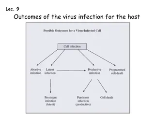
Three potential outcome of EBV infection 1. EBV can replicate in B cells or epithelial cells permissive for EBV replication (acute phase). 2. EBV can cause latent infection of B cells in the presence of competent T cell immunity (healthy carrier stage). 3. EBV can stimulate and immortalize B cells. 4. Implicate in several cancers.
Infectious Mononuclosis Primary EBV infection is usually subclinical in childhood. However in adolescents and adults, there is a 50% chance that the syndrome of infectious mononucleosis (IM) will develop. IM is usually a self-limited disease which consists of fever, lymphadenopathy and splenomegaly. In some patients jaundice may be seen which is due to hepatitis. Atypical lymphocytes are present in the blood. Complications occur rarely but may be serious e.g. splenic rupture, meningoencephalitis, and pharyngeal obstruction. In some patients, chronic IM may occur where eventually the patient dies of lymphoproliferative disease or lymphoma. Diagnosis of IM is usually made by the heterophil antibody test (Monospot Test) and/or detection of anti- EBV VCA IgM. There is no specific treatment.
Disease Association 1. Infectious Mononucleosis 2. Burkitt's lymphoma 3. Nasopharyngeal carcinoma 4. Lymphoproliferative disease and lymphoma in the immunosuppressed. 5. X-linked lymphoproliferative syndrome 6. Chronic infectious mononucleosis 7. Oral leukoplakia in AIDS patients 8. Chronic interstitial pneumonitis in AIDS patients.
Laboratory Diagnosis The diagnosis of infectious mononucleosis in the clinical laboratory is based primarily on two approaches: 1.In the hematologic approach, absolute lymphocytosis occurs and as many as 30% abnormal lymphocytes are seen on a smear. These atypical lymphs are large and have a lobulated nucleus and a vacuolated, basophilic cytoplasm. They are cytotoxic T cells that are reacting against the EBV-infected B cells. 2.In the immunologic approach, there are two types of serologic tests: (a) The heterophil antibody test is useful for the early diagnosis of infectious mononucleosis because it is usually positive by week 2 of illness. However, because the antibody titer declines after recovery, it is not useful for detection of prior infection. The Monospot test is often used to detect the heterophil antibody; it is more sensitive, more specific, and less expensive than the tube agglutination test. (b) The EBV-specific antibody tests are used primarily in diagnostically difficult cases. The IgM VCA antibody response can be used to detect early illness; the IgG VCA antibody response can be used to detect prior infection. In certain instances, antibodies to EA and EBNA can be useful diagnostically.
Cytomegalovirus Normal individuals Primary infection is usually asymptomatic, occasionally an infectious mononucleosis-like illness may be seen. Reactivations or re-infections are common throughout life and are usually asymptomatic.
Epidemiology CMV is one of the most successful human pathogens, it can be transmitted vertically or horizontally usually with little effect on the host. Transmission may occur in utero, perinatally or postnatally. Once infected, the person carries the virus for life which may be activated from time to time, during which infectious virions appear in the urine and the saliva. Reactivation can also lead to vertical transmission. It is also possible for people who have experienced primary infection to be reinfected with another or the same strain of CMV, this reinfection does not differ clinically from reactivation. In developed countries with a high standard of hygiene, 40% of adolescents are infected and ultimately 70% of the population is infected. In developing countries, over 90% of people are ultimately infected.
Clinical Manifestations Congenital infection - may result in cytomegalic inclusion disease Perinatal infection - usually asymptomatic Postnatal infection - usually asymptomatic. However, in a minority of cases, the syndrome of infectious mononucleosis may develop which consists of fever, lymphadenopathy, and splenomegaly. The heterophil antibody test is negative although atypical lymphocytes may be found in the blood. Immunocompromised patients such as transplant recipients and AIDS patients are prone to severe CMV disease such as pneumonitis, retinitis, colitis, and encephalopathy. Reactivation or reinfection with CMV is usually asymptomatic immunocompromised patients. except in
Congenital Infection Defined as the isolation of CMV from the saliva or urine within 3 weeks of birth. Commonest congenital viral infection, affects 0.3 - 1% of all live births. The second most common cause of mental handicap after Down's syndrome and is responsible for more cases of congenital damage than rubella. Transmission to the fetus may occur following primary or recurrent CMV infection. 40% chance of transmission to the fetus following a primary infection. May be transmitted to the fetus during all stages of pregnancy. No evidence of teratogenecity, damage to the fetus results from destruction of target cells once they are formed.
Cytomegalic Inclusion Disease CNS abnormalities - microcephaly, mental retardation, spasticity, epilepsy, periventricular calcification. Eye - choroidoretinitis and optic atrophy Ear - sensorineural deafness Liver - hepatosplenomegaly and jaundice which is due to hepatitis. Lung - pneumonitis Heart - myocarditis Thrombocytopenic purpura, Haemolytic anaemia Late sequelae in individuals asymptomatic at birth - hearing defects and reduced intelligence.
Laboratory Diagnosis (1) In general, the detection of CMV from blood specimens or bronchioalveolar lavage is more prognostic of clinical CMV disease than the detection of the virus from urine or saliva. 1. Virus isolation (a) Conventional cell culture - human embryo lung fibroblasts used, requires 1 to 3 weeks for characteristic CPE to appear, remains the gold standard for the diagnosis of CMV infection (b) Rapid culture methods (Shell-vial technique)- e.g. DEAFF test - detects the expression of CMV early antigens within 24 to 48 hours of inoculation. Appears to be as sensitive and specific as conventional cell culture.
Laboratory Diagnosis (2) 2. CMV antigenaemia test - widely used in many European countries. CMV polymorphonuclear leukocytes are detected by immunoperoxidase or immunofluorescence techniques. A result can be obtained within 4 to 6 hours but the technique is very tricky. The pp65 CMV antigenaemia test is now routinely used for the rapid diagnosis of CMV infection in immunocompromised patients. antigens at the surface of 3. Polymerase chain reaction - becoming the method of choice in a few laboratories, had been reported to carry a higher prognostic value for CMV disease than the DEAFF test. 4. Serology - not reliable in general but occasionally, rises in IgG titre and the presence of IgM may be seen.
Prevention No licensed vaccine is available. Prevention of CMV disease in transplant recipients is a very complicated subject and varies from center to center. It may include the following measures. Screening and matching the CMV status of the donor and recipient Use of CMV negative blood for transfusions Administration of CMV immunoglobulin to seronegative recipients prior to transplant ganciclovir Give prophylactically. antiviral agents such as
HHV6 and HHV7 Isolated from AIDS patients. HHV6 is the causative agent of roseola infantum or exanthem subitum ; HHV7 remains an orphan virus. Lymphotropic (target CD4+lymphocytes). The main target cell is the T-lymphocyte, although B- lymphocytes may also be infected. Asymptomatic to mild infections universal in human populations. Roseola infantum or exanthem subitum, a common childhood disease that resolves spontaneously. It is thought that HHV-6 and HHV-7 are related to each other in a similar manner to HSV-1 and HSV-2.
Epidemiology and Pathogenesis HHV-6 and HHV-7 are ubiquitous and are found worldwide. They are transmitted mainly through contact with saliva and through breast feeding. HHV-6 and HHV-7 infection are acquired rapidly after the age of 4 months when the effect of maternal antibody wears off. By the time of adulthood, 90-99% of the population had been infected by both viruses. Like other herpesviruses, HHV-6 and HHV-7 remains latent in the body after primary infection and reactivates from time to time.
Human Herpes Virus 8 the gammaherpesviruses Belong herpesviruses Originally isolated from cells of Kaposi s sarcoma (KS) Now appears to be firmly associated with Kaposi s sarcoma as well as some lesser known malignancies such as Castleman s disease and primary effusion lymphomas to subfamily of HHV-8 DNA is found in almost 100% of cases of Kaposi s sarcoma Most patients with KS have antibodies against HHV-8 The seroprevalence of HHV-8 is low among the general population but is high in groups of individuals susceptible to KS, such as homosexuals
In 1994, it was reported that a new herpesvirus, now known as HHV-8, or Kaposi's sarcoma associated herpesvirus (KSHV), may be the cause of Kaposi's sarcoma (KS), the most common cancer in patients with AIDS. The idea that a virus other than HIV is the cause of KS arose from epidemiologic data that showed that KS was common in patients who acquired HIV sexually but rare in patients who acquired HIV via blood transfusion. A second virus transmitted sexually appeared likely to be the cause.
HHV-8 causes malignant transformation by a mechanism similar to that of other DNA viruses (such as human papillomavirus), namely, inactivation of a tumor suppressor gene. A protein encoded by HHV-8 called nuclear antigen inactivates the RB (retinoblastoma) tumor suppressor protein, which causes the cells to grow in an uncontrolled manner. Transmission of HHV-8 is primarily sexually, but it is also transmitted in transplanted organs such as kidneys and appears to be the cause of transplantation-associated KS. The DNA of HHV-8 is found in the cells of transplantation-associated KS but not in the cells of other transplantation-associated cancers. KS in AIDS patients is a malignancy of vascular endothelial cells that contains many spindle-shaped cells and erythrocytes. The lesions are dark purple, flat to nodular, and often appear at multiple sites such as the skin, oral cavity, and soles (but not the palms). Internally, lesions occur commonly in the gastrointestinal tract and the lungs. The extravasated red cells give the lesions their purplish color. HHV- 8 also infects B cells, inducing them to proliferate and produce a type of lymphoma called primary effusion lymphoma.
Laboratory diagnosis of KS is often made by biopsy of the skin lesions. HHV-8 DNA and RNA are present in most spindle cells, but that analysis is not usually done. Virus is not grown in culture. The type of treatment depends upon the site and number of the lesions. Surgical excision, radiation, and systemic drugs, such as alpha interferon or vinblastine, can be used. There is no specific antiviral therapy and no vaccine against HHV-8.
")


")


")