Esophageal Surgery and Dysfunction
Learn about esophageal surgery, the structure of the esophagus, causes of dysfunction, and general surgical principles to follow. Discover the complexities of treating issues in this vital connecting tube between the pharynx and stomach.
Download Presentation

Please find below an Image/Link to download the presentation.
The content on the website is provided AS IS for your information and personal use only. It may not be sold, licensed, or shared on other websites without obtaining consent from the author. If you encounter any issues during the download, it is possible that the publisher has removed the file from their server.
You are allowed to download the files provided on this website for personal or commercial use, subject to the condition that they are used lawfully. All files are the property of their respective owners.
The content on the website is provided AS IS for your information and personal use only. It may not be sold, licensed, or shared on other websites without obtaining consent from the author.
E N D
Presentation Transcript
Esophageal surgery Dr. Rafid Majeed
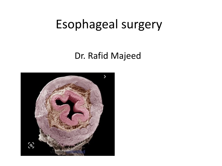
The esophagus is the connecting tube between the pharynx and stomach that functions to transport ingesta and fluids. Esophagus The outer layer of the esophagus is the adventitia. In the neck, the esophageal adventitia blends with the deep cervical fascia. In the thorax and abdomen, the adventitia is largely covered with pleura and peritoneum, respectively. The esophagus is loosely connected to the diaphragm by a phrenicoabdominal membrane. The muscularis is composed of striated muscle for the entire length of the esophagus in dogs, but it changes to smooth muscle in the terminal esophagus in cats. The submucosa loosely connects the mucosa and muscularis, allowing mucosa to move independently and form mucosal folds in the undistended esophagus. The submucosa contains blood vessels, nerves, and simple mucous glands that secrete mucus, which lubricates the mucosal surface. The esophageal mucosa is composed of a stratified squamous epithelium. In the nondistended esophagus, the mucosa forms numerous large longitudinal folds that can be seen with positive-contrast esophagography. In cats, the terminal esophagus is also folded transversely
Causes of dysfunction of the esophageal phase of swallowing can be broadly divided into 1. mechanical (or anatomic) lesions (a luminal or mural lesion or compression from adjacent structures like foreign bodies or tumors ) 2. functional (or neuromuscular) lesions (neuromuscular disease and commonly causes megaesophagus) 3. inflammatory (esophagitis) conditions (include ingestion of corrosive substances, thermal burns, radiation injury, and esophageal foreign bodies.)
GENERAL SURGICAL PRINCIPALS Esophageal surgery is historically associated with a higher prevalence of incisional dehiscence than surgery on other portions of the alimentary tract. Several factors may contribute to the high complication rate, including 1. lack of serosa, 2. the segmental nature of the blood supply, 3. the lack of omentum, 4. constant motion caused by swallowing and respiration, 5. tension at the surgical site esophageal surgery can be successfully performed by following principles, including gentle tissue handling, minimization of contamination, appropriate selection and application of suture materials, appropriate use of electrocautery, and accurate apposition of tissues
In the abdomen, the serosa assists in healing of viscera by the elaboration of a fibrin seal soon after surgery and by providing a source of pluripotential mesothelial cells. In the thoracic cavity, pleural mesothelium may play a similar role. The blood supply to the esophagus is considered segmental, with a rich, intramural plexus of anastomosing vessels in the submucosal layer that can support long segments of the esophagus. Postoperative motion of the esophageal incision with swallowing and respiration is unavoidable. After esophageal surgery, food and water are normally withheld to reduce esophageal motion and eliminate the passing of ingesta across the surgical site.
DISEASES OF THE ESOPHAGUS Vascular Ring Anomalies Vascular rings are developmental anomalies of the great vessels that result in the encircling of esophagus and trachea by a complete or incomplete ring of vessels. Several types of vascular ring anomalies are described in dogs and cats. The most common vascular ring anomaly is persistent right aortic arch with a left ligamentum arteriosum, in which the aortic arch develops from the right fourth aortic arch . The clinical signs of vascular ring anomalies are caused primarily by esophageal obstruction.. Initially, regurgitation usually occurs soon after eating, but later it may occur at variable time (minutes to hours).
Treatment Most vascular ring anomalies can be corrected through a left lateral thoracotomy. The ligamentum arteriosum is carefully dissected from the esophagus with right-angled forceps double ligated with silk suture, and transected. Prognosis with surgical correction of persistent right aortic arch with left ligamentum arteriosum in dogs has improved over the past several decades.
Congenital Generalized Megaesophagus Idiopathic congenital total megaesophagus is caused by generalized alteration in motor function of the esophagus, possibly from a defect in vagal afferent innervation of the esophagus, and results in lack of aboral propulsion of food The entire esophagus becomes dilated, and esophagitis may develop as a result of fermentation of retained food. Treatment : Esophagodiaphragmatic cardioplasty using the Torres technique to improve esophageal emptying.
Esophagodiaphragmatic cardioplasty The esophagus was approached through a ninth left intercostal thoracotomy and isolated with moistened saline sponges. The vagus nerves were identified and avoided. The diaphragm was dissected free of the left half of the esophagus at the esophageal hiatus, and a semicircular shaped segment of diaphragmatic muscle (2 to 3 cm wide) was resected. The new diaphragmatic edge was sutured to the esophagus with full- thickness horizontal mattress sutures of 2-0 polypropylene. A final layer of sutures (3-0 absorbable monofilament) was placed to close any remaining gaps in the diaphragm cardioplasty places light tension on the distal esophagus; with contraction and relaxation of the diaphragm during respiration, the cardia also underwent intermittent contraction and dilatation, resulting in a pumping mechanism that helped drain the esophagus. Prognosis : good
Esophageal Foreign Bodies Esophageal foreign bodies are a common problem in dogs and are occasionally diagnosed in cats. The most common foreign bodies in dogs are ingested bones. In cats, fishhooks, needles, and string foreign bodies are more common. Small-breed dogs, particularly terrier breeds, are most frequently diagnosed with esophageal foreign bodies. Most affected dogs are younger than 3 years of age (64%) Acute clinical signs associated with complete or severe partial obstruction are usually seen. The most classic clinical sign is regurgitation of food within a few minutes of eating. Water is generally retained unless there is complete obstruction. Other clinical signs include retching , gagging, excessive salivation, restlessness, lethargy , and inappetence Chronically affected animals may remain bright and alert but have weight loss and periodic bouts of regurgitation and inappetence. Sharp or chronic foreign bodies can result in esophageal perforation,
Treatment An initial attempt should be made to extract the foreign body with endoscopy using grasping forceps. the firmly lodged foreign body should not be forced because this may induce or enlarge a perforation. Then Surgical repair of perforations should be considered . Esophageal foreign bodies should be surgically removed when retrieval or advancement of the foreign body fails or when forceps extraction presents a significant risk for laceration of the esophagus or major vessels Prognosis The prognosis after foreign body removal is generally excellent except in cases of thoracic esophageal perforation
Esophageal Lacerations Esophageal lacerations are most commonly caused by penetrating esophageal foreign bodies. The most common injury is a penetrating stick injury in dogs that carry, chew, or retrieve sticks. Esophageal stick injuries generally occur in the cervical esophagus. Esophageal lacerations can also occur with external trauma such as bite wounds Treatment Foreign material is removed surgicaly, the esophageal laceration is minimally debrided, and a single- or double layered closure performed. The surgical site is lavaged before closure, and a closed suction drain can be placed. Acute esophageal penetrating injuries have a poorer prognosis
Esophageal Diverticula Esophageal diverticula are rare in dogs and cats. diverticula are divided into pulsion and traction types . A pulsion diverticulum is an outpouching of mucosa that herniates through a defect in the tunica muscularis. It is thought to be caused by increased luminal pressure as a result of a mechanical (e.g., foreign body or stricture) or functional esophageal obstruction. A traction diverticulum is a full-thickness deviation of the esophageal wall. The term traction refers to the assumed pathogenesis, namely inflammation in an adjacent organ causing formation of an adhesion. Subsequent contraction of the adhesion pulls the esophagus outward to form a pouch The diverticulum can become impacted with ingesta, distorting and obstructing the esophageal lumen
Treatment Small diverticula may be treated conservatively by feeding a gruel-type diet with the animal in an upright position Large diverticula generally require surgical management. Excision of the diverticulum (diverticulectomy) Prognosis The prognosis in dogs with diverticula amenable to simple diverticulectomy is good
Esophageal Fistulae An esophageal fistula is an abnormal communication between the esophagus and the trachea, bronchus, lung parenchyma, or (less commonly) the skin. Esophageal fistulae may be either congenital or acquired. Congenital fistulae result from an incomplete separation of the tracheobronchial tree from the digestive tract most acquired fistulae are secondary to esophageal foreign bodies The most common clinical sign is coughing, which may be associated with drinking liquids. Other clinical signs include regurgitation, lethargy, anorexia, pyrexia, dyspnea, and weight loss.
Treatment and Prognosis Esophageal fistulae require surgical management. The esophageal defect can usually be closed primarily, although more extensive resection and reconstruction techniques may be required.
Esophageal Neoplasia Esophageal neoplasia in dogs and cats is rare. In dogs and cats, the most commonly reported primary esophageal tumors include squamous cell carcinoma, Clinical Signs The most common clinical sign is regurgitation or vomiting
