Esophageal Varices: Anatomy, Physiology, and Management
Esophageal varices are abnormal, enlarged veins in the lower esophagus often linked to liver diseases. Explore the anatomy, physiology, incidence, etiology, symptoms, diagnosis, and management of this condition.

Download Presentation

Please find below an Image/Link to download the presentation.
The content on the website is provided AS IS for your information and personal use only. It may not be sold, licensed, or shared on other websites without obtaining consent from the author.If you encounter any issues during the download, it is possible that the publisher has removed the file from their server.
You are allowed to download the files provided on this website for personal or commercial use, subject to the condition that they are used lawfully. All files are the property of their respective owners.
The content on the website is provided AS IS for your information and personal use only. It may not be sold, licensed, or shared on other websites without obtaining consent from the author.
E N D
Presentation Transcript
PRESENTED BY:- MR.SHAILENDRA VERMA NURSING TUTOR RCN, BAREILLY
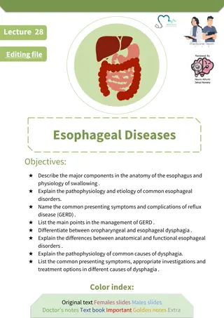
SPECIFIC OBJECTIVES SPECIFIC OBJECTIVES On the completion of class teaching the group will be able to:- Introduce the topic Define the Oesophageal varices Explain the anatomy and physiology Explain the incidence of Oesophageal varices Enlist the Etiology of Oesophageal varices Describe the pathophysiology of Oesophageal varices Explain the Sign and Symptoms Oesophageal varices Explain the diagnostic evaluation of Oesophageal varices Describe the Management of Oesophageal varices Enlist Complications of Oesophageal varices
INTRODUCTION INTRODUCTION It is disorder of GI. Esophageal varices are abnormal, enlarged veins in the tube that connects the throat and stomach (esophagus). This condition occurs most often in people with serious liver diseases.
DEFINITION DEFINITION Esophageal varices (Sometimes spelled esophageal varix or oesophageal varices) are extremely dilated sub-mucosal veins in the lower third of the esophagus. They are most often a consequence of portal hypertension, commonly due to cirrhosis; people with esophageal varices have a strong tendency to develop bleeding
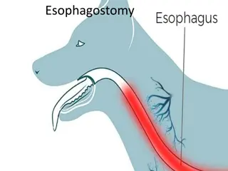
ANATOMY AND PHYSIOLOGY OF ANATOMY AND PHYSIOLOGY OF OESOPHAGEAL OBSTRUCTION OESOPHAGEAL OBSTRUCTION
INCIDENCE INCIDENCE It is more common in males than in females. 50 % of patients with esophageal varices will experience
ETIOLOGY / RISK FACTORS ETIOLOGY / RISK FACTORS Causes of esophageal varices include: Severe liver scarring (cirrhosis). Blood clot (thrombosis). A blood clot in the portal vein or in a vein that feeds into the portal vein (splenic vein) can cause esophageal varices. A parasitic infection. The parasite can damage the liver, as well as the lungs, intestine and bladder
High portal vein pressure. (portal hypertension). Large varices. The larger the varices, the more likely they are to bleed. Red marks on the varices. Severe cirrhosis or liver failure. Continued alcohol use.
PATHOPHYSIOLOGY PATHOPHYSIOLOGY Due to etiological factors Portal venous hypertension Increased resistance to flow in portal venous system Increased pressure Portal systemic shunting (abnormal venous communication between portal system and systemic venous circulation) Appearing a large submucosal vein at lower end of esophagus and gastric fundus Hemmorhage due to increased intravariceal pressure Esophageal varices
CLINICAL MANIFESTATION CLINICAL MANIFESTATION Signs and symptoms can include: Vomiting and seeing significant amounts of blood in your vomit Black, tarry or bloody stools Light-headedness Loss of consciousness (in severe case) Yellow coloration of your skin and eyes (jaundice) Easy bleeding or bruising Fluid buildup in your abdomen (ascites)
DIAGNOSTIC EVALUATION DIAGNOSTIC EVALUATION History collection & physical examination Blood investigation (if needed) Endoscope exam. Imaging tests. Capsule endoscopy. In this test, you swallow a vitamin-sized capsule containing a tiny camera, which takes pictures of the esophagus as it goes through your digestive tract. This might be an option for people who are unable or unwilling to have an endoscope exam. This technology is more expensive than regular endoscopy and not as available.
MANAGEMENT MANAGEMENT Medications to reduce pressure in the portal vein. A type of blood pressure drug called a beta blocker may help reduce blood pressure in your portal vein, decreasing the likelihood of bleeding. These medications include propranolol (Inderal, Innopran) and nadolol (Corgard).
Surgical Management Surgical Management Using elastic bands to tie off bleeding veins. If your esophageal varices appear to have a high risk of bleeding, your doctor might recommend a procedure called band ligation. Using an endoscope, the doctor snares the varices and wraps them with an elastic band, which essentially "strangles" the veins so they can't bleed. Esophageal band ligation carries a small risk of complications, such as scarring of the esophagus
NURSING ASSESSMENT: NURSING ASSESSMENT: Assessment ABCD Initial acute Airway Does the patient have a patent airway? Are they at significant risk of losing their airway? Breathing Respiratory rate, oximetry, respiratory effort, air entry? Circulation Heart rate, blood pressure, central and peripheral capillary refill time? IV access? How much blood is the patient losing? Disability: Level of consciousness? Are they distressed? Irritable (consider encephalopathy)? Are they in pain? Are they febrile? Receiving antibiotics?
PREVENTION PREVENTION Don't drink alcohol. Eat a healthy diet. Maintain a healthy weight. Use chemicals sparingly and carefully. Reduce your risk of hepatitis.
COMPLICATIONS COMPLICATIONS Rebleeding, Shock, which can lead to Death,
SUMMARY SUMMARY After completed my teaching session group improve their knowledge, skills in identifying patient with oesophageal obstruction, and also apply in your future regarding:- Introduce the topic Define the Oesophageal varices Explain the anatomy and physiology Explain the incidence of Oesophageal varices Enlist the Etiology of Oesophageal varices Describe the pathophysiology of Oesophageal varices Explain the Sign and Symptoms Oesophageal varices Explain the diagnostic evaluation of Oesophageal varices Describe the Management of Oesophageal varices Enlist Complications of Oesophageal varices
RECAPTUALIZATION RECAPTUALIZATION Define Oesophageal varices Cause of Oesophageal varices Explain pathophysiology of varices Describe the management of varices
ASSIGNMENT / APPLICATION : ASSIGNMENT / APPLICATION :- - Assignment on NCP of pre and post surgical management. Date of Submission ---------------
REFERENCES REFERENCES Lippincott Williams and Wilkins, text book of Manual of Nursing Practice Ninth Ed, publisher by wolters kluwer, New Delhi page no 685-687 Arlene L. Polaski and Suzanne E. Tatro, text book of Luckmann s Core Principle of Medical Surgical 2010 , publisher by Elsevier, India page no 484- 487 Brunners And Suddharths, 2010, Text book of Medical Surgical Nursing, 12thedition ,volume 2, publisher by wolters kluwer , page no 104-107 Linda S. Williams and Paula.D Hopper ,2003, Understanding Medical Surgical Nursing , 2ndEd, FA Davis publisher, page no 836-838 BT Basavanthappa, text book of Medical Surgical Nursing 2ndEd, publisher by Jaypee Brothers, page no 520-524