Evolution of Carious Lesion Classification
Carious lesion classification has evolved over time, incorporating new concepts such as the understanding of fluoride's action and the importance of adhesive restorative materials. These advancements have led to a paradigm shift in dentistry towards prevention and limiting cavity size, challenging previous limitations in classification systems like G.V. Black's criteria. Clinical examination techniques have also improved, emphasizing the use of sharp explorers and blunt probes to minimize enamel damage. This modern approach enhances treatment precision and outcomes.
Download Presentation

Please find below an Image/Link to download the presentation.
The content on the website is provided AS IS for your information and personal use only. It may not be sold, licensed, or shared on other websites without obtaining consent from the author.If you encounter any issues during the download, it is possible that the publisher has removed the file from their server.
You are allowed to download the files provided on this website for personal or commercial use, subject to the condition that they are used lawfully. All files are the property of their respective owners.
The content on the website is provided AS IS for your information and personal use only. It may not be sold, licensed, or shared on other websites without obtaining consent from the author.
E N D
Presentation Transcript
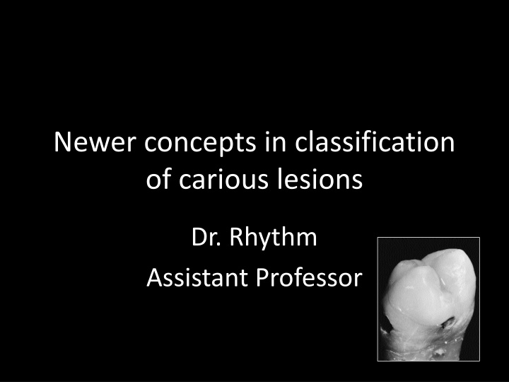
Newer concepts in classification of carious lesions Dr. Rhythm Assistant Professor
Earlier, cavities were designed without the present understanding of the action of the fluoride ion and, in the presence of restorative materials which had no inherent therapeutic properties, were subject to micro leakage and were often not aesthetic.
In the absence of adhesive restorative materials, it was regarded as essential to remove all unsupported enamel regardless of its location.
MACRO DENTISTRY G.V BLACKS EXTENSION FOR PREVENTION MICRO DENTISRY PREVENTION OF EXTENSION
In the presence of a better understanding of the caries process and improved knowledge of the function of fluoride, it is now possible to limit the size of a cavity
CLINICAL EXAMINATION CLINICAL EXAMINATION G.V.Black a sharp explorer should be used with some pressure & if a very slight pull is required to remove it i.e. CATCH POINT , the pit should be marked for restoration even if there are no signs of decay .
SHARP EYES SHARP EYES BUT BUT BLUNT PROBE BLUNT PROBE The enamel is damaged by forceful probing with sharp sickle probes, so probes used to examine occlusal surfaces should be blunt and the probing forces light
Limitations of Blacks Classification The five categories of carious lesion were related to the site of the lesion and to the nature of the intended restoration but they did not take into account the dimensions of a cavity
Blacks criterion 1) To remove tooth structure to gain access andvisibility 2) To remove all traces of affected dentine from the floor of the cavity 3) To make room for the insertion of the restorative material itself
4) To provide a mechanical interlocking retentive designs, and 5) To extend the cavity to self cleansing areas to avoid recurrent caries.
Classification (Mount & Hume, 1998) Size Minimal Moderate Enlarged Extensive 1 2 3 4 Site Pit/fissure 1 1.1 1.2 1.3 1.4 Contact area 2 2.1 2.2 2.3 2.4 Cervical 3 3.1 3.2 3.3 3.4 Australian Dental Journal 1998;43:3.
Three sites of carious lesions Site 1. Pits, fissures and enamel defects on occlusal surfaces of posterior teeth or other smooth surfaces, such as cingulum pits on anteriors.
Site 2. Approximal enamel immediately below areas in contact with adjacent teeth.
Site 3. The cervical one-third of the crown or, following gingival recession, the exposed root.
4 sizes of the lesion Size 1. Minimal involvement of dentine just beyond treatment by re-mineralization alone. Size 2. Moderate involvement of dentine. Size 3. The cavity is enlarged beyond moderate. Size 4. Extensive caries with bulk loss of tooth structure has already occurred.
The Size 1 cavity will necessarily be a new lesion and adhesive restorative materials are ideal, and should always be used for restoration under these circumstances
Cavities in the Size 2, 3 and 4 range may be new lesions that have progressed to a considerable extent without the patient presenting for treatment or they may result from a breakdown of an old restoration which requires replacement.
Black Class II Site 2, Size 2 (#2.2) in both posteriors and anteriors.
Black Class III Site 2, Size 2 (#2.2) and extends to Site 2, Size 3 (#2.3) Black Class IV Site 2, Size 4 (#2.4)
Black Class V An erosion/abrasion lesion or a small carious cavity would be a Site 3, Size 1 (#3.1) or Site 3, Size 2 (#3.2) and interproximal lesions would generally be recorded as Site 3, Size 3(#3.3) or Site 3, Size 4 (#3.4).
MCQ Q1 The new classification is based on A. frequency of lesion B. shape of lesion C. site of lesion D. depth of lesion
Q. 2 Blacks principles are based on A. extension and prevention B. extension for prevention C. prevention of extension D. extensive prevention.
Q.3 Site 1 lesion corresponds to A. pit and fissure caries B. smooth surface lesions C. erosive defects D. anterior tooth caries
Q.4 Site 2 lesions correspond to A. Blacks class III B. Black s class I C. Black s class V D. Black s class VI
Q. 5 Caries on exposed proximal root surface are classified under A. Site 3 B. Site 2 C. Site 1 D. Not classified.
