Ex Vivo Analysis of Leishmania Infection in Human Blood Cells
A study conducted by Dr. Medhavi Ranatunga, investigating how various human blood cell populations interact with Leishmania parasites to establish infection over 24 hours. Results show infection percentages in monocytes, neutrophils, and B cells, with higher rates in neutrophils and monocytes. The study sheds light on the roles of different cell types in Leishmania infection.

Download Presentation

Please find below an Image/Link to download the presentation.
The content on the website is provided AS IS for your information and personal use only. It may not be sold, licensed, or shared on other websites without obtaining consent from the author.If you encounter any issues during the download, it is possible that the publisher has removed the file from their server.
You are allowed to download the files provided on this website for personal or commercial use, subject to the condition that they are used lawfully. All files are the property of their respective owners.
The content on the website is provided AS IS for your information and personal use only. It may not be sold, licensed, or shared on other websites without obtaining consent from the author.
E N D
Presentation Transcript
Ex vivo analysis of Leishmania infection in human blood cells over 24 hours Dr Medhavi Ranatunga Postdoctoral Academic in the Biosciences M.Ranatunga@greenwich.ac.uk
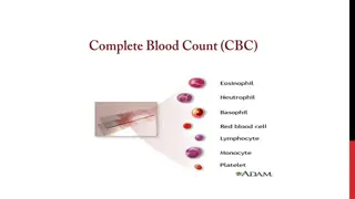
Introduction A number of human cells are thought to be able to host Leishmania parasites but their effect/participation on establishment of infection remains widely uncharacterized. Neutrophil Monocyte Macrophage Dendritic cell B lymphocyte
Aims To investigate how whole blood cell populations participate and interact with Leishmania parasites to allow establishment of infection in humans.
Material and Methods Red Blood cell lysed Whole Blood Immunophenotyping Macrophage Neutrophil Anti- CD16b Anti-CD20 Anti- CD14 4h & 24h Alexa Fluor CD14 PE-Cy CD20 PE CD16b CD16b CD20 CD14 Monocyte Dendritic cell Lymphocytes GFP expressing Metacyclic Promastigotes Flow cytometry
Results and Discussion (Percentage of infection) Monocyte (M ) Neutrophil (PMN) B Lymphocyte (B cells) CD16B - CD16B + CD14 - CD20 - CD14 + CD20 + Monocyte (M ) B Lymphocyte (B cells) Neutrophil (PMN) CD16B - CD14 - CD16B + CD20 - CD14 + CD20 + Percentage of infection in M Percentage of infection in B cells Percentage of infection in PMN Percentage of infection Percentage of infection Percentage of infection 4H 24H 24H 4H 24H 24H 4H 24H 24H 4H 4H 4H L. aethiopica L. mexicana L. aethiopica L. mexicana L. aethiopica L. mexicana
Conclusion During whole blood infection both L. aethiopica and L. mexicana infect Neutrophils, Monocytes and B cells over 24 hours. As expected higher percentage of Neutrophils and Monocytes get infected when comparing to B cells during whole blood infection.
Reference 1. Getti, G. T et al. (2008). Induction of apoptosis in host cells: a survival mechanism for Leishmania parasites?. Parasitology. 135, 1391-1399. Ruben, E et al. (2012). In Vitro and In Vivo Efficacy of Ether Lipid Edelfosine against Leishmania spp. and SbV-Resistant Parasites. PLos Neglected Tropical Diseases. 6 (4), 1-14. Liu D, Uzonna JE. The early interaction of Leishmania with macrophages and dendritic cells and its influence on the host immune response. Front Cell Infect Microbiol [Internet]. 2012;2(June):83. Available from: http://journal.frontiersin.org/article/10.3389/fcimb.2012.00083/ab stract DaMata JP, Mendes BP, Maciel-Lima K, Menezes CAS, Dutra WO, Sousa LP, et al. Distinct macrophage fates after in vitro infection with different species of Leishmania: Induction of apoptosis by Leishmania (Leishmania) amazonensis, but not by Leishmania (Viannia) guyanensis. PLoS One [Internet]. 2015;10(10). Available from: http://dx.doi.org/10.1371/journal.pone.0141196 2. 3. 4.
Thank you ! Dr. Giulia Getti Dr. Paul Dyer Rachel Nice Atiya Raza Samantha Lewis Samantha Ingram