Exploring Globular Proteins: Structure, Functions & Examples
This content delves into the world of globular proteins, focusing on their structure, functions, and key examples like hemoglobin and myoglobin. Discover how these proteins fold into spherical shapes, enhancing their solubility, and learn about their role in oxygen transport, storage, immune function, and enzymatic reactions. Gain insights into different types of hemoglobin, abnormal variants, and diseases associated with globular proteins.

Download Presentation

Please find below an Image/Link to download the presentation.
The content on the website is provided AS IS for your information and personal use only. It may not be sold, licensed, or shared on other websites without obtaining consent from the author. If you encounter any issues during the download, it is possible that the publisher has removed the file from their server.
You are allowed to download the files provided on this website for personal or commercial use, subject to the condition that they are used lawfully. All files are the property of their respective owners.
The content on the website is provided AS IS for your information and personal use only. It may not be sold, licensed, or shared on other websites without obtaining consent from the author.
E N D
Presentation Transcript
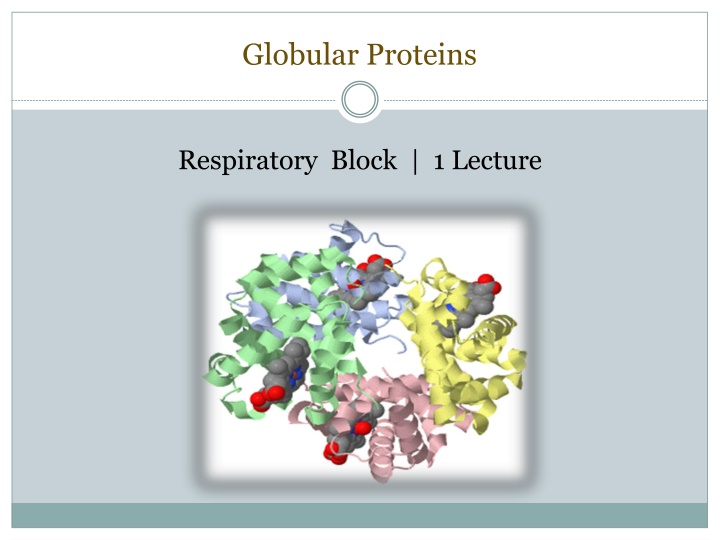
Globular Proteins Respiratory Block | 1 Lecture
Objectives To describe the globular proteins using common examples like hemoglobin and myoglobin. To study the structure and functions of globular proteins like- Hemoglobin (a major globular protein) Myoglobin, and -globulins (immunoglobulins) To know the different types of hemoglobin and difference between normal and abnormal hemoglobin To understand the diseases associated with globular proteins
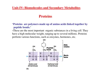
Globular proteins Amino acid chains fold into shapes that resemble spheres are called globular proteins This type of folding increases solubility of proteins in water Polar groups on the protein s surface Hydrophobic groups in the interior Fibrous proteins are structural proteins mainly insoluble
Globular proteins Hemoglobin: oxygen transport function Myoglobin: oxygen storage/supply function in heart and muscle 1, 2, -globulins: various functions -globulins (immunoglobulins): immune function Enzymes: catalysis of biochemical reactions
Hemoglobin A major globular protein in humans Composed of four polypeptide chains: Two and two chains Contains two dimers of subunits Held together by non-covalent interactions Each chain is a subunit with a heme group in the center that carries oxygen A Hb molecule contains 4 heme groups and carries 4 molecules of O2
Types of Hb Normal: HbA (97%) HbA2 (2%) HbF (1%) HbA1c Carboxy Hb Met Hb Sulf Hb Abnormal:
HbA structure Oxygen binding to hemoglobin
Hemoglobin function Carries oxygen from the lungs to tissues Carries carbon dioxide from tissues back to the lungs Normal level (g/dL): Males: 14-16 Females: 13-15
Types of hemoglobin Fetal hemoglobin (HbF): Major hemoglobin found in the fetus and newborn Tetramer with two and two chains Higher affinity for O2 than HbA Transfers O2 from maternal to fetal circulation across placenta
Types of hemoglobin HbA2: Appears ~12 weeks after birth Constitutes ~2% of total Hb Composed of two and two globin chains
Types of hemoglobin HbA1c: HbA undergoes non- enzymatic glycosylation Glycosylation depends on plasma glucose levels HbA1c levels are high in patients with diabetes mellitus NH2 groups of the N-terminal valines of chains
Abnormal Hbs Unable to transport O2 due to abnormal structure: Carboxy-Hb: CO replaces O2 and binds 200X tighter than O2 (in smokers) Met-Hb: Contains oxidized Fe3+ (~2%) that cannot carry O2 Sulf-HB: Forms due to high sulfur levels in blood (irreversible reaction)
Hemoglobinopathies Disorders of hemoglobin caused by: Synthesis of structurally abnormal Hb Synthesis of insufficient quantities of normal Hb Combination of both
Hemoglobinopathies Sickle cell (HbS) disease Caused by a single mutation in -globin gene Glutamic acid at position 6 in HbA is replaced by valine The mutant HbS contains s chain The shape of RBCs become sickled Causes sickle cell anemia
Hemoglobinopathies Hemoglobin C disease: Caused by a single mutation in -globin gene Glutamic acid at position 6 in HbA is replaced by lysine Causes a mild form of hemolytic anemia
Hemoglobinopathies Methemoglobinemia: Caused by oxidation of Hb to ferric (Fe3+) state Methemoglobin cannot bind oxygen Caused by certain drugs, reactive oxygen species and NADH-cytochrome b5 reductase deficiency Chocolate cyanosis: brownish-blue color of the skin and blood
Hemoglobinopathies Thalassemia: Defective synthesis of either or -globin chain due to gene mutation -thalassemia: Synthesis of -globin chain is decreased or absent Causes mild to moderate hemolytic anemia -thalassemia: Synthesis of -globin chain is decreased or absent Causes severe anemia Patients need regular blood transfusions
Myoglobin A globular hemeprotein in heart and muscle Stores and supplies oxygen to the heart and muscle only Contains a single polypeptide chain forming a single subunit with eight -helix structures The interior of the subunit is composed of nonpolar amino acids
Myoglobin The charged amino acids are located on the surface The heme group is present at the center of the molecule Myoglobin gives red color to skeletal muscles Supplies oxygen during aerobic exercise
Myoglobin in disease Myoglobinuria: Myoglobin is excreted in urine due to muscle damage (rhabdomyolysis) May cause acute renal failure Specific marker for muscle injury Less specific marker for heart attack
Immunoglobulins Defensive proteins produced by the B-cells of the immune system Y-shaped structure with 2 heavy and 2 light polypeptide chains Neutralize bacteria and viruses Types: IgA, IgD, IgE, IgG, IgM
Take Home Messages Amino acid chains fold into shapes that resemble spheres are called globular proteins. Fibrous proteins are mainly insoluble, while globular proteins are soluble structural proteins. Hb, Myoglobin, globulines and enzymes are examples of globular proteins. Functionally, Hb is for O2 and CO2 transport. HbA, HbA2 and HbF are examples of normal Hb, in which the tetrameric structure is composed of 2 constant subunits with 2 changeable subunits according to Hb type.
Take Home Messages HbA1C is a HbA which undergoes non-enzymatic glycosylation, depending on plasma glucose levels. Carboxy-Hb, Met-Hb and Sulf-Hb are examples of abnormal Hb, in which O2 molecules are not transported due to abnormal Hb structure. Disorders of Hb caused by synthesis of structurally abnormal Hb and/or insufficient quantities of normal Hb. Sickle cell (HbS) and HbC diseases are caused by a single mutation in -globin gene.
Take Home Messages Glu6 in HbS is replaced by Val, while it is replaced by Lys in HbC. Methemoglobinemia is caused by oxidation of Hb, inhibiting O2 binding leading to chocolate cyanosis. Thalassemia is caused by a defect in synthesis of either - or -globulin chain, as a result of gene mutation. -Thalassemia causes less sever anemia than - Thalassemia. Myoglobin is a globular hemeprotein, which stores and supplies O2 to the heart and muscle only.
Take Home Messages Hb is composed of 4 chains (subunits), while Myoglobin is composed of a single chain. Myoglobinuria is a specific marker for muscle injury and may cause acute renal failure. Immunoglobulins are defensive proteins produced by the B-cells. Immunoglobulins consist of 5 types: IgA, IgD, IgE, IgG and IgM.
References Illustrations in Biochemistry by Lippincott 6th edition.