Factor Deficiencies and Coagulation Abnormalities in Pediatrics
This content explores factor deficiencies and coagulation abnormalities in infants and children, covering the physiology of hemostasis, pathophysiology of Hemophilia, and perioperative management. It delves into primary hemostasis, platelet responses, and the coagulation cascade, providing valuable insights for medical professionals.

Uploaded on Feb 20, 2025 | 2 Views
Download Presentation

Please find below an Image/Link to download the presentation.
The content on the website is provided AS IS for your information and personal use only. It may not be sold, licensed, or shared on other websites without obtaining consent from the author.If you encounter any issues during the download, it is possible that the publisher has removed the file from their server.
You are allowed to download the files provided on this website for personal or commercial use, subject to the condition that they are used lawfully. All files are the property of their respective owners.
The content on the website is provided AS IS for your information and personal use only. It may not be sold, licensed, or shared on other websites without obtaining consent from the author.
E N D
Presentation Transcript
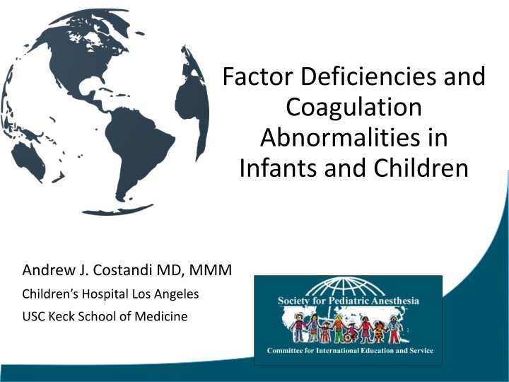
Factor Deficiencies and Coagulation Abnormalities in Infants and Children Andrew J. Costandi MD, MMM Children s Hospital Los Angeles USC Keck School of Medicine
Faculty Disclosures None
Learning Objectives Upon completion of this activity, participants will be able to: Describe the physiology of hemostasis in the pediatric patient Describe the pathophysiology of various types of Hemophilia Discuss appropriate perioperative management of children with Hemophilia.
Hemostasis Primary Hemostasis Vascular spasm Platelet plug formation Secondary Hemostasis Coagulation Cascade Formation of blood clot Tertiary Hemostasis Clot lysis Vascular remodeling
Primary Hemostasis Platelet Response 1. Platelet adhesion 2. Platelet activation 3. Platelet aggregation "Blood clotting" by Alexey Kashpersky, Radius Digital Science is licensed under CC BY-NC 4.0
Primary Hemostasis Platelet Response 1. Platelet Adhesion Normal oEndothelial cells lining the vascular wall exhibit antithrombotic properties Intimal injury oRelease of procoagulant subendothelial elements like collagen and VWF Platelet adhesion to site of injury oCollagen on the subendothelial surface is bound by platelet integrins oVWF on the subendothelial surface is bound by platelet glycoprotein GPIb-IX-V
Primary Hemostasis Platelet Response 2. Platelet Activation Release of oThromboxane A2 oFibrinogen oFactor V oADP Conformational and shape change oElongated pseudopods oExtremely adhesive platelets
Primary Hemostasis Platelet Response 3. Platelet Aggregation o Thromboxane A2 and ADP stimulate platelet aggregation o VWF and fibrinogen bridge between platelets o Coagulation cascade is triggered and fibrin is deposited. "Blood clotting" by Alexey Kashpersky, Radius Digital Science is licensed under CC BY-NC 4.0
Secondary Hemostasis The Classic model By Dr Graham Beards - Own work, CC BY-SA 3.0, https://commons.wikimedia.org/w/index.php?curid=19094276
Cell-based Model of Coagulation Simultaneous, interactive pathways that overlap to augment production of thrombin Termination Propagation Initiation Amplification
Secondary Hemostasis 1. Extrinsic X-ase 3.Intrinsic X-ase 2. Prothrombinase Joe D [CC BY-SA 3.0 (http://creativecommons.org/licenses/by-sa/3.0/)] 4. Protein C Complex
Tertiary Hemostasis Tissue plasminogen activators activate plasminogen to plasmin Fibrinolysis
Neonatal Coagulation ~ 50% adult levels at birth o Decreased Vitamin K dependent factors (II, VII, IX, X) o Decreased factor XI and XII, Prekallikrein, Kallikrein o Decreased anticoagulant proteins (C, S, AT III) Vitamin K critical for coagulation Neonates have low Vitamin K
Bleeding Disorders In Children Coagulation Protein Disorders Acquired Disorders: Vitamin K Deficiency Bleeding Liver Disease Chronic Kidney Disease Coagulation Inhibitors Disseminated intravascular coagulation (DIC)
Bleeding Disorders In Children Coagulation Protein Disorders Inherited coagulation protein disorders Rare (3-5% of congenital bleeding disorders) Autosomal recessive Isolated factor deficiency: o Hemophilia (VIII, IX) o Other (I, II, V, VII, X, XI)
Clinical Presentation Purpuric Dysfunction (disorders of platelets and blood vessels) Bleeding into skin and mucous membranes Petechiae Small ecchymoses Excessive bleeding after minor trauma or surgery Coagulation Protein Disorder Large ecchymoses Hemarthrosis Soft tissue hematomas Excessive bleeding after surgery
Laboratory Tests Initial Testing CBC and platelets Peripheral blood smear Coagulation Studies o PTT (INR) o aPTT o Fibrinogen
Laboratory Tests Platelet Count Disorder PT/INR aPTT Fibrinogen Qualitative Platelet Disorder Normal/Low Normal Normal Normal Quantitative Platelet Disorder Low Normal Normal Normal Hemophilia Normal Normal Prolonged Normal Von Willebrand Disease (VWD) Normal Normal Normal/Prolonged Normal DIC Low Prolonged Prolonged Low
Coagulation Protein Disorders Hemophilia Isolated Factor Deficiency: o Fibrinogen, o Factor VII o Factor V o Factor II o Factor X o Factor XIII
Hemophilia X linked bleeding disorder 1 in 10,000 births
Types of Hemophilia (based on factor deficiency) Hemophilia A Hemophilia B Hemophilia C Acquired Hemophilia +/- Congenital Hemophilia with Inhibitors ? CC0 1.0 Universal (CC0 1.0) Public Domain
Hemophilia A Mutation in the long arm of chromosome X at F8 gene Deficiency or lack of factor VIII (FVIII) 80 85% of the total hemophilia population 1 in 5000 males 20-30 % develop inhibitory antibodies CC0 1.0 Universal (CC0 1.0) Public Domain
Hemophilia B Christmas Disease Mutation in the long arm of chromosome X at F9 gene Deficiency or lack of coagulation factor IX (FIX) 1 in 25,000 males 1-6% develop inhibitory antibodies CC0 1.0 Universal (CC0 1.0) Public Domain
Hemophilia C Factor XI deficiency Homozygotes: < 4% factor XI Heterozygotes: 15-65% factor XI Autosomal recessive Higher Incidence in Ashkenazi Jewish population Bleeding diathesis may not correlate well with factor concentrations
Acquired Hemophilia Rare Potentially life-threatening bleeding disorder Autoantibodies against endogenous plasma coagulation factors
Congenital Hemophilia with Inhibitors Development of IgG Ab against EXOGENEOUS factor 10-20% with severe hemophilia A 1-5% with severe hemophilia B Median age of 3 years Suspected when an increase in the frequency of bleeding occurs
Congenital Hemophilia with Inhibitors Bethesda units (BU) = amount of inhibitors present 1 BU = 50% inactivation of: factor VIII or IX in 1 mL of plasma Positive for inhibition = > 0.6 BU/mL High responder >5BU/mL
Hemophilia: Clinical Presentation Mild and Moderate Deficiency o Bleeding after trauma or surgery Severe Deficiency: o Spontaneous bleeding Serious Life threatening Joints (hemarthrosis) Muscles (deep compartments calf and forearm) Mucous membranes in the mouth, gums, nose, and genitourinary tract Intracranial Neck/throat Gastrointestinal
Hemophilia: Clinical Presentation Severe Hemophilia Clinical Presentation Presents with spontaneous bleeding in the first two years of life. Common sites of bleeding by age: o Newborn Central Nervous System Sites of medical interventions: circumcision, heel stick o Toddler: Frenulum Oral injury o Children: Bruising Joint bleed o Older children and Adults: Hemarthrosis (80% of hemorrhages)
Hemophilia: Classification Factor Activity Age at Diagnosis Severity Symptoms Frequent spontaneous bleeding; abnormal bleeding after minor injuries, surgery, or tooth extractions Severe < 1% Age 2 years Rare spontaneous bleeding; abnormal bleeding after minor injuries, surgery, or tooth extractions Moderate 1% - 5% Age <5-6 years No spontaneous bleeding; abnormal bleeding after major injuries, surgery, or tooth extractions Often later in life, depending on hemostatic challenges Mild > 5% - 40%
Hemophilia: Diagnosis Bleeding profile: Hemophilia Prolonged bleeding time Normal platelet count Normal PT Prolonged or normal aPTT Establish diagnosis Factor assay (FVIII, FIX)
Hemophilia: Treatment Guidelines for perioperative factor levels in patients with hemophilia for major and minor surgical procedures. Source: Srivstava A, Brewer AK, Mauser-Bunschoten et al. Haemophilia. 2013 Jan;19:e1-47.
Hemophilia A: Treatment MILD MODERATE Desmopressin (DDAVP) SEVERE Factor Replacement Viral inactivated plasma-derived Recombinant factor concentrates Cryoprecipitate Fresh Frozen Plasma SEVERE WITH INHIBITORS Factor Replacement Bypassing Agents "DDAVP" by ballookey is licensed under CC BY-NC-ND 2.0
Desmopressin (DDAVP) Protects factor VIII from degradation Presents Factor VIII to site of bleeding Produces 3-5 fold increase in VWF:FVIII Peak @ 30-90 mins Duration of 8-12 hours Intranasal dose = 150 mcg < 50 kg or 300 mcg > 50 kg IV dose = 0.3 mcg/kg (max 20mcg) "DDAVP" by ballookey is licensed under CC BY-NC-ND 2.0
Desmopressin (DDAVP) Mild or Moderate hemophilia A No value in hemophilia B Low cost No risk of transmission of viral infections Complications of prolonged treatment: o Tachyphylaxis "DDAVP" by ballookey is licensed under CC BY-NC-ND 2.0 o Hyponatremia
Hemophilia B: Treatment MILD, MODERATE & SEVERE Factor Replacement Fresh Frozen Plasma "DDAVP" by ballookey is licensed under CC BY-NC-ND 2.0
Factor VIII Concentrates Recombinant FVIII (Recombinate) o Number of units of FVIII required = Weight of patient x % factor level desired x 0.5 o 1 Unit/Kg IV will raise the plasma FVIII level by 2% o Half time FVIII: 8 12 hours Plasma derived factor VIII (Monoclate P & Hemofil-M) Plasma derived factor VIII-VWF (Humate P) Porcine factor VIII concentrates
Factor IX Concentrates Recombinant Factor IX Concentrate (BeneFIX) o Number of units of FVIII required = Weight of patient x % factor level desired x 1 o 1 Unit/Kg IV will raise the plasma FVIII level by 1% o Half time FIX: 18 24 hours Plasma-derived Factor IX Concentrate (Alphanite) Prothrombin complex concentrates (PCCs) o Contains factors II, VII, IX and X
Cryoprecipitate or FFP? Cryoprecipitate 1 ml = 3-5 U FVIII + VWF + Fibrinogen + FXIII, but not FIX or FXI Risk of viral pathogen transmission Fresh frozen plasma Large volume needed Risk of viral pathogen transmission Limited rise of FVIII levels
Clot Stabilizers Antifibrinolytics Promotes clot stability by inhibiting the conversion of plasminogen to plasmin -> inhibiting fibrinolysis Adjunctive therapy in VWD and Hemophilia Valuable in oral and dental surgery Can cause nausea and vomiting Contraindicated in patients treated with PCC
Clot Stabilizers: Antifibrinolytics Tranexamic acid (TXA) 10 mg/kg IV q8h 25 mg/kg PO daily Epsilon aminocaproic acid 50 mg/kg IV or PO Shorter plasma half-life Less potent than TXA CC BY-SA 4.0
Hemophilia with Inhibitors Low Titers/Low Responders: Defined as Inhibitor level < 5 BU/ml High Dose Factor Replacement Porcine Factor VIII High Titers/High Responders Defined as Inhibitor level 5 BU/ml Bypassing Agent (rFVIIa and aPCC) Public domain.
Hemophilia With Inhibitors: Treatment High-titer/High-responding inhibitors: Bypassing Agents Preoperative dosing Postoperative dosing Days 1 5 Days 6 14 Recombinant Factor VIIa (rFVIIa) 90 120 g/kg q2h up to four times then q3 6 h for 24 h Minor Surgery 120-150 g/kg q 2h 90 120 g/kg q2 h Day 1, then q3 h Day 2, then q4 h Days 3 5 90-120 g/kg q6 h Intermediate/Major Surgery 150 g/kg q 2h Continuous infusion 15 50 g/kg/h 15-50 g/kg/h 15 50 g/kg q 1h Activated Prothrombin Complex Concentrate (aPCC) 50 75 U/kg q12 24 h one to two times Minor Surgery 50 75 U/kg 75 100 U/kg q12 h Intermediate/Major Surgery 75 100 U/kg 75 100 U/kg q8 12 h Kulkarni R1. Comprehensive care of the patient with haemophilia and inhibitors undergoing surgery: practical aspects. Haemophilia. 2013 Jan;19(1):2-10. doi: 10.1111/j.1365-2516.2012.02922.x. Epub 2012 Aug 27.
Suggested Reading Smith SA. The cell-based model of coagulation. J Vet Emerg Crit Care. 2009;19(1):3-10. Lee, JW. Von Willebrand Disease, Hemophilia A and B, and Other Factor Deficiencies. Int Anesthesiol Clin. 2004;42(3):59-76. Srivastava A, Brewer AK, Mauser-Bunshoten EP, et al. Guidelines for the management of hemophilia. Hemophilia. 2013;19(1):e1-47. Srivastava A. Hemophilia care beyond the treatment guidelines. Hemophilia. 2014; Suppl 4:4-10. Elsey NM. Hemophilias. In: Lalwani K, Cohen IT, Choi EY, Raman VT, ed. Pediatric Anesthesia: A Problem-Based Learning Approach. Oxford University Press; 2018 Kulkani R. Comprehensive care of the patient with haemophilia and inhibitors undergoing surgery: practical aspects. Hemophilia. 2013;19(1):2-10. doi: 10.1111/j.1365-2516.2012.02922.x. Epub 2012 Aug 27.


")
")