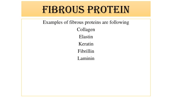
Fibrous protein
Fibrous proteins like collagen, elastin, and keratin are essential for structural functions in the body. Collagen and elastin, common examples, play a vital role in skin, connective tissues, and blood vessels. The unique structure of fibrous proteins, derived from specific amino acids, provides them with special mechanical properties. Learn about the types of collagen and the distinct structures they form in various organs. The amino acid sequence of collagen, rich in proline and glycine, is crucial for forming the characteristic triple-stranded helix structure.

Download Presentation

Please find below an Image/Link to download the presentation.
The content on the website is provided AS IS for your information and personal use only. It may not be sold, licensed, or shared on other websites without obtaining consent from the author.If you encounter any issues during the download, it is possible that the publisher has removed the file from their server.
You are allowed to download the files provided on this website for personal or commercial use, subject to the condition that they are used lawfully. All files are the property of their respective owners.
The content on the website is provided AS IS for your information and personal use only. It may not be sold, licensed, or shared on other websites without obtaining consent from the author.
E N D
Presentation Transcript
Fibrous protein Examples of fibrous proteins are following Collagen Elastin Keratin Fibrillin Laminin
Collagen and elastin, examples of common, well- characterized fibrous proteins of the extracellular matrix that serve structural functions in the body. Collagen and elastin, found as components of skin, connective tissue, blood vessel walls, and sclera and cornea of the eye. Fibrous protein exhibits special mechanical properties, resulting from its unique structure, which are obtained by combining specific amino acids into regular, secondary structural elements. Fibrous proteins provide tensile streng This is totally different from globular proteins, whose shapes are the result of complex interactions between secondary, tertiary, and quaternary structural elements.
Types of collagen There are 25 types of collagens, some examples are as following- Major collagen present in bone - Type I(90%) Major collagen present in dermis ,ligaments and tendons -Type I (80%) Major collagen present in cartilage - type II (40-50%) Major collagen present in aorta - type I and type III (20-40%) Major collagen present in basement membrane type IV Major collagen present in hemi desmose type XVII
Structure of collagen A typical collagen molecule --- long, rigid structure in which three polypeptides (referred to as chains ) are wound around one another in a rope-like triple helix Although these molecules are found throughout the body, their types and organization are dictated by the structural role collagen plays in a particular organ. Gel like structure: Vitreous humor Tight parallel structure: Tendon Stacked transparent structure: Cornea Arranged at an angle: Bone
Amino acid sequence: - Collagen is rich in proline and glycine Both of which are important in the formation of the triple-stranded helix. Proline facilitates the formation of the helical conformation of each chain because its ring structure causes kinks in the peptide chain Presence of proline dictates that the helical conformation of the chain cannot be an helix.(don t confused with alpha helix)) Glycine, the smallest amino acid, is found in every third position of the polypeptide chain. The glycine residues are part of a repeating sequence, Gly X Y , where X is frequently proline and Y is often hydroxyproline (but can be hydroxylysine) Most of the chain can be regarded as a polytripeptide whose sequence can be represented as ( Gly Pro Hyp )
Quarter staggered arrangement Lateral association of triple helical units Each is displaced longitudinally from its neighbor by slightly less than one quarter of its length Above two factors are responsible for tensile strength of collagen fibers
Synthesis of collagen Intracelluler event Takes place inside the rough endoplasmic reticulum of fibroblast-formation of peptide chains of procollagen Cleavage of signal peptide by proteolytic enzymes Hydroxylation of prolyl residues and some lysyl residues Glycosylation of some hydroxylysyl residues Formation of intrachain and interchain S-S bonds in extension peptides Formation of triple helix Procollagen is formed
Extracellular events Takes place in extracellular matrix Cleavage of amino and carboxyl terminal propeptides Assembly of collagen fibres in quarter staggered alignment Oxidative deamination of amino groups of lysyl and hydroxylysyl residues to aldehydes Formation of intra and interchain covalent cross-links via Schiff bases and aldol condensation products Tropocollagen is formed
Unique events in collagen formation occurs both in intracellular and extracellular 1.Hydroxylation Post translational modification occurring intracellularly Enzyme- prolyl and lysyl hydroxylase Coenzyme vitamin C and alpha ketogluterate Fe is cofactor and O2 is needed for hydroxylation reaction Essential for three chains of monomer to fold into triple helix at body temperature (hydroxylation provides cross linkages between the alpha chains of collagen) In case of vitamin C deficiency which is coenzyme of hydroxylation defective connective tissues of bones are formed which further lead to weak bone
2.Glycosylation Intracellular event Hydroxylysine residues are glycosylated with galactose and glucose
3.Oxidative deamination Extracellular event Enzyme lysyl oxidase Cofactor copper Oxidative deamination of lysyl and hydroxylysyl residues to form aldehydes 4.Covalent crosslinks After oxidative deamination modified lysyl and hydroxylysyl residues forms covalent cross links by aldol condensation Provides tensile strength to collagen
Applied aspects In next slide there is a table showing different diseases due to defective or deficient different types of collagen Just go through
Type of collagen Diseases Type I Osteogenesis imperfecta Osteoporosis Ehlers-Danlos Syndrome Chondrodysplasias Osteoarthritis Ehler-Danlos syndrome Type II Type III Type IV Alport syndrome Type V and Type I Classical Ehler-Danlos syndrome Type III Hypermobile EDS Type VII Epidermolysis bullosa Type X Schmid metaphyseal chondrodysplasia Lysyl hydroxylase Ehler-Danlos syndrome Lysyl oxidase Menkes disease
Alport syndrome X linked disorder Type IV collagen is affected Clinical features hematuria ,sensorineural deafness,conical deformation of anterior surface of lens
Achondroplasia Most common cause of chondrodysplasia Most common cause of short limb dwarfism Caused by mutation in gene that codes for the receptor for fibroblast growth factor -3
ELASTIN A connective tissue that is responsible for Properties of extensibility and elastic recoil in tissues Present in lung, large arteries blood vessels, and some elastic ligaments
UNIQUE FEATURES OF ELASTIN Oxidative deamination and desmosine cross links in elastin Enzyme lysyl oxidase Lysyl residues of tropoelastin are oxidatively deaminated to aldehydes. Condensation of three of these lysine derived aldehydes with an unmodified lysine to form a tetra functional cross-link unique to elastin
Clinical correlation of elastin Mutations in elastin gene leads to supravalvular aortic stenosis (William Beuren Syndrome) Cutis laxa
Fibrillin Glycoprotein Structural component of micro fibrils Secreted into the extracellular matrix by fibroblast Incorporated into the insoluble micro fibrils Provide a scaffold for deposition of elastin Applied aspect:Mutation in gene (on chromosome 15)for fibrillin 1 causes Marfan syndrome Tried of Marfan syndrome are- 1.Skeletal changes abnormal periosteum 2. Ectopia lentis abnormal lens due to abnormal zonular fibers 3.Aortic aneurysm
Keratin Two alpha helix are wind around one another to form super helix Keratin is rich in hydrophobic amino acids like alanine ,leucine,methionine,valine ,phenylalanine Crosslinks are formed by disulphide bonds When more disulphide bonds are present more harder the keratin is Keratin is present in hair ,nail and outer layer of skin Mutations in the genes for major keratin of basal epithelial cells causes a clinical condition Epidermolysis bullosa simplex
Laminin Major protein component of renal glomerular and other basal laminas It has elongated cruciform shape Basal lamina has three fibrous proteins-laminin ,entactin and type IV collagen as well as Hboparin and heparin sulphate glycosaminoglycans