fMRI Preprocessing Techniques
Explore the necessity of preprocessing in fMRI, covering topics like realignment, unwarping, violations of key assumptions, causes of head movement, and motion prevention strategies in this informative visual presentation.

Download Presentation

Please find below an Image/Link to download the presentation.
The content on the website is provided AS IS for your information and personal use only. It may not be sold, licensed, or shared on other websites without obtaining consent from the author. If you encounter any issues during the download, it is possible that the publisher has removed the file from their server.
You are allowed to download the files provided on this website for personal or commercial use, subject to the condition that they are used lawfully. All files are the property of their respective owners.
The content on the website is provided AS IS for your information and personal use only. It may not be sold, licensed, or shared on other websites without obtaining consent from the author.
E N D
Presentation Transcript
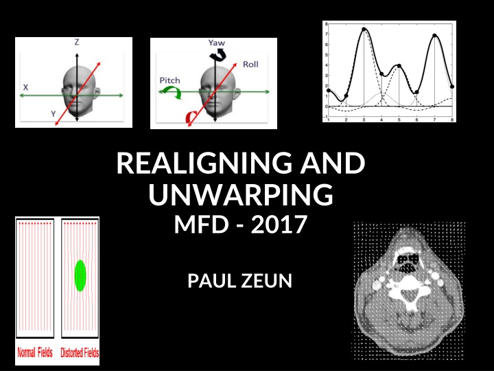
REALIGNING AND UNWARPING MFD - 2017 PAUL ZEUN
What this talk covers Preprocessing in fMRI : Why is it needed? Motion in fMRI Realignment Unwarping How this all works in SPM
Stages in fMRI analysis Scanner Output Preprocessing Statistical analysis Today s talk Motion correction (and unwarping) Design matrix fMRI time series General Linear Model Smoothing Statistical Parameter Map Spatial normalisation (including coregistration) Structural MRI Parameter estimates
Preprocessing: Why is it needed? fMRI: returns a 3D array of voxels repeatedly sampled over time Changes in activation in each voxel correlated with experimental task Key Assumptions: 1) all voxels must be acquired simultaneously 2) the voxels need to come from the same part of the brain
Violation of assumption 1: - all voxels must be acquired simultaneously - the last slice is acquired TR seconds after the first slice there can be motion of one slice relative to another Solution: - slice-timing correction: include realignment parameters in the model - will (hopefully) be discussed in event-related fMRI
Violation of assumption 2: - the voxels need to come from the same part of the brain - small movement (< 5mm) means that voxel location is not stable throughout the time series Voxel A - Inactive Voxel A - Active
Causes of head movement: - Actual movement of the head - Physiological: heart beat, respiration, blinking - Task-related: moving to press buttons - artificially creates variance in voxel activation that correlates with task serious confound Why is this a problem? - this variance is often much larger than experiment- induced variance False activations - lowers the signal-to-noise ratio
Motion Prevention in fMRI 1. Constrain the volunteer s head (soft padding) 2. Give explicit instructions to lie as still as possible, not to talk between sessions, and swallow as little as possible 3. Try not to scan for too long* 4. Make sure your subject is as comfortable
Realignment Realignment involves two stages: 1. Registration Estimate the 6 parameters that describe the rigid body transformation between each image and a reference image 2. Transformation Re-sample each image according to the determined transformation parameters
Realignment: Rigid body transformation To correct for both rotation and translation, we will compute 6 parameters Translation Rotation Yaw Roll Pitch Translations about Z axis about Y axis about X axis cos( ) 0 sin( ) 0 sin( ) 0 cos( ) 0 cos( ) sin( ) 0 0 sin( ) cos( ) 0 0 1 0 0 0 0 1 0 0 0 0 1 0 Xtrans Ytrans Ztrans 1 1 0 0 0 0 0 0 0 0 1 0 1 0 0 0 0 0 1 0 0 1 0 0 0 0 1 cos( ) sin( ) 0 sin( ) cos( ) 0
Optimisation - cost function - quantifies how (dis-)similar the images are after a spatial transformation has been applied - Minimise sum of squared difference Least squares between consecutive images
Realigning: 2. Interpolation Original image Resampled image After having realigned, we need to determine the intensity of each new voxel Realign 1. Original voxels 2. New voxels to determine after realigning 3. For example, want to determine this voxel 4. 3 types of interpolation possible: 1. Nearest Neighbour 2. Trilinear 3. B-Spline Original voxel New voxel to identify
Simple Interpolation Nearest neighbour Takes the value of the closest voxel Tri-linear Weighted average of the neighbouring voxels * f5 = f1 x2 + f2 x1 * f6 = f3 x2 + f4 x1 * f7 = f5 y2 + f6 y1
B-spline Interpolation A continuous function is represented by a linear combination of basis functions
Realigning: Head movement Calculate position of the brain for the 1st slice Estimate transformation parameters based on 1st slice Apply the transformation parameters on each slice
Summary so far Motion can reduce sensitivity + increase residual variance Try to minimise movement with practical steps Motion correction via realignment - Each volume rigidly registered to reference - Least squares objective function Realigned images must be resliced (interpolation) for analysis
Residual Errors in Realigned fMRI Even after realignment a considerable amount of the variance can be accounted for by effects of movement This can be caused by e.g.: 1. Movement between and within slice acquisition 2. Re-sampling can introduce Interpolation errors 3. Non-linear distortions and drop-out due to inhomogeneity of the magnetic field Incorporate movement parameters as confounds in the statistical model
Registration A. realignment = registration that uses a linear transformation that preserves shape B. unwarping = registration using non-linear transformation that modifies shape
Unwrap tackles non-linear distortion from magnetic field inhomogeneities (movement by inhomogeneity interaction) 1) Different substances in the brain are differentially magnetised susceptible to magnetization 2) Inhomogeneity of the magnetic field 3) Distortion of the image
Field Maps Measure field inhomogeneity using a field map Using this map derive a deformation field (how voxels in volume are deflected due to distorted field)
Measure deformation field (FieldMap). Estimate new distortion fields for each image Unwarp time series Estimate rate of change of field with respect to current estimate of pitch & roll Estimate movement parameters + B 0 B 0
Practicalities of unwarping Particularly useful when the movements correlate with activation of interest as can remove unwanted variance without removing true activations. Can dramatically reduce variance in areas susceptible to greatest distortion (e.g. orbitofrontal cortex and regions of the temporal lobe). Useful when high field strength or long readout time increases amount of distortion in images
Summary Importance of motion in fMRI Corrected by using realignment and rigidly registering each volume to a reference Then reslice interpolate images to match reference voxels Movement by distortion interaction corrected by unwarping
References SPM 12 Manual - www.fil.ion.ucl.ac.uk/spm/doc/manual.pdf SPM course slides - www.fil.ion.ucl.ac.uk/spm/course/ Previous MfD slides