Functions of Cerebral Hemisphere and Cortex
The cerebral hemispheres are the largest part of the brain, with distinct functions and structures such as the cerebral cortex, lobes, and cortical regions. The cortex consists of six layers, each playing a specific role in sensory processing and motor functions. Primary motor and sensory areas, as well as association areas, are crucial for processing signals and coordinating responses within the brain.

Download Presentation

Please find below an Image/Link to download the presentation.
The content on the website is provided AS IS for your information and personal use only. It may not be sold, licensed, or shared on other websites without obtaining consent from the author.If you encounter any issues during the download, it is possible that the publisher has removed the file from their server.
You are allowed to download the files provided on this website for personal or commercial use, subject to the condition that they are used lawfully. All files are the property of their respective owners.
The content on the website is provided AS IS for your information and personal use only. It may not be sold, licensed, or shared on other websites without obtaining consent from the author.
E N D
Presentation Transcript
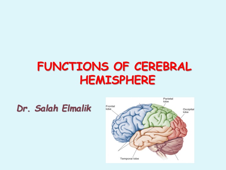
FUNCTIONS OF CEREBRAL HEMISPHERE Dr. Salah Elmalik
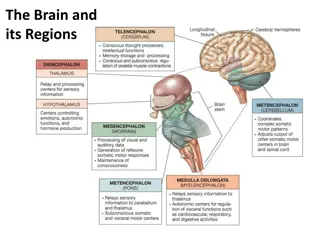
The Cerebrum Lobes, the Cerebral Cortex, and Cortical Regions of the Brain
The cerebrum is the largest part of the brain with two hemisphere, linked by commissural fibres of corpus callosum. Each cerebral hemisphere contains externally highly convoluted cortex of grey matter and internal mass of white matter or medulla. The cerebral hemispheres contains motor and sensory areas and the limbic system.
Cerebral Cortex - The outermost layer of gray matter making up the superficial aspect of the cerebrum. Cerebral Cortex CerebralCortex
Microscopically the cortex consists of six layers or laminae lying parallel to the surface From outside to inside the layers are: I. Molecular layer II. The external granular layer III. Layer of pyramidal cell IV. Internal granular layer V. large pyramidal cell layer VI. Layer of fusiform or polymorphic cells.
The incoming sensory signal excites neuronal layer IV first; then the signal spreads toward the surface of the cortex and also toward deeper layers. 2.Layers I and II & III perform most of intracortical association function. 3.The neurons in layers II and III making short horizontal connections with adjacent cortical areas. 4.The neurons in layers V and VI send output signals to brain stem ,spinal cord (V) & thalamus (VI)
Primary Areas: The primary motor areas have direct connections with specific muscles for causing discrete muscle movements. The primary sensory areas detect specific sensations visual, auditory, or somatic transmitted directly to the brain from peripheral sensory organs. Association Areas They receive and analyze signals simultaneously from multiple regions of both the motor and sensory cortices as well as from subcortical structures.
These areas receive and analyze signals simultaneously from multiple regions of both the motor and sensory cortices as well as from subcortical structures. The most important association areas are (1) Parieto-occipitotemporal association area (2) prefrontal association area (3) limbic association area.
1. Analysis of the Spatial Coordinates of the Body. 2. Language Comprehension. 3. Initial Processing of Visual Language (Reading). 4. Area for Naming Objects.
AREA SITE FUNCTION computes the coordinates of the visual , auditory, and body surroundings. Analysis of the Spatial Coordinates of the Body. beginning in the posterior parietal cortex and extending into the superior occipital cortex Wernicke's area, lies behind the primary auditory cortex in the posterior part of the superior gyrus of the temporal lobe angular gyrus area Area for Language Comprehension higher intellectual function Area for Initial Processing of Visual Language (Reading). make meaning out of the visually perceived words (lesion causes Dyslexia or Word Blindness) naming objects. Area for Naming Objects. Lateral portion of anterior occipital lobe & posterior temporal
Is the anterior pole of frontal lobe. It contributes in the following functions: 1. Planning of complex pattern of movements. 2. Personality characteristics and social relationship 3. Production of deep, more abstract and logically sequenced thoughts which enable attainment of goals 4. Working memory (ability to tie thoughts together in a logical sequence by comparing many bits of information with appropriate stored knowledge and be able to instantly recall this information for future planning) Lesions in this area lead to change in personality and behavior
Consists of anterior and inner portion of temporal lobe. Is primarily concerned with emotion , behavior and motivational drive for different tasks most importantly learning. Lesion of this area may lead to decreased aggression , lack of emotion , hyper sexuality & hyperphagia
Located on the underside of the brain on the medial occipital and temporal lobes The occipital portion is contiguous with visual cortex , while the temporal one is closely associated with limbic system Inability to recognize faces is called prosopagnosia
Frontal Parietal Occipital Temporal
The Frontal Lobe of the brain is located deep to the Frontal Bone of the skull It plays an integral role in the following functions: - Memory Formation -Emotions -Decision Making Reasoning - Personality
Primary Motor Cortex (Precentral Gyrus) Cortical site involved with controlling movements of the body. Broca s Area plan of motor pattern for expressing of individual words. Located on Left Frontal Lobe. Broca s Aphasia Results in the ability to comprehend speech, but the decreased motor ability (or inability) to speak and form words. Orbitofrontal Cortex Site of Frontal Lobotomies * Possible Side Effects: - Epilepsy - Poor Emotional Responses - Perseveration (Uncontrolled, repetitive actions, gestures, or words) * Desired Effects: - Diminished Rage - Decreased Aggression - Poor Emotional Responses Olfactory Bulb - Cranial Nerve I, Responsible for sensation of Smell
Primary Motor Cortex/ Precentral Gyrus Broca s Area Orbitofrontal Cortex Olfactory Bulb Regions Regions
The Parietal Lobe of the brain is located deep to the Parietal Bone of the skull. It plays a major role in the following functions/actions: - Senses and integrates sensation(s) - Spatial awareness and perception (Proprioception - Awareness of body/ body parts in space and in relation to each other)
Primary Somatosensory Cortex (Postcentral Gyrus) Site involved with processing of tactile and proprioceptive information. Somatosensory Association Cortex - Assists with the integration and interpretation of sensations relative to body position and orientation in space. May assist with visuo-motor coordination. Primary Gustatory Cortex Primary site involved with the interpretation of the sensation of Taste.
Parietal lobe is essential for our feeling of touch, warmth/heat , cold, pain , body position and appreciation of shapes of palpated objects . When damaged , the person loses the ability to recognize shapes of complex objects by palpation (palpation = examination of objects by touch ) & develops Sensory Inattention on opposite side 22
Primary Somatosensory Cortex/ Postcentral Gyrus Somatosensory Association Cortex Primary Gustatory Cortex Regions Regions
The Occipital Lobe of the Brain is located deep to the Occipital Bone of the Skull. Its primary function is the processing, integration, interpretation, etc. of VISION and visual stimuli.
Primary Visual Cortex This is the primary area of the brain responsible for sight - recognition of size, color, light, motion, dimensions, etc. Visual Association Area Interprets information acquired through the primary visual cortex.
Primary Visual Cortex Visual Association Area
The Temporal Lobes are located on the sides of the brain, deep to the Temporal Bones of the skull. They play an integral role in the following functions: - Hearing -Organization/Comprehensi onof language - Information Retrieval (Memory and Memory Formation)
Primary Auditory Cortex Responsible for hearing Primary Olfactory Cortex Interprets the sense of smell once it reaches the cortex via the olfactory bulbs. (Not visible on the superficial cortex) Wernicke s Area Language comprehension. Located on the Left Temporal Lobe.
Lesion may lead to: - - Wernicke s Aphasia Language comprehension is inhibited. Words and sentences are not clearly understood, and sentence formation may be inhibited or non-sensical. Memory impairment & can be associated with temporal lobe epilepsy 29
Primary Auditory Cortex Wernike s Area Primary Olfactory Cortex (Deep) Conducted from Olfactory Bulb Regions Regions
Each cerebral hemisphere receives sensory information from, and sends motor commands to, the opposite side of body The 2 hemispheres have somewhat different functions although their structures are alike Correspondence between a specific function and a specific region of cerebral cortex is not precise No functional area acts alone; conscious behavior involves the entire cortex 1. 2. 3. 4.
Functional differences between left and right hemispheres In most people, left hemisphere (dominant hemisphere) controls: reading, writing, and math, decision-making, logic, speech and language (usually) Right cerebral hemisphere relates to: understanding & interpreting music, Non verbal visual Experience Spatial relation between the person & their surroundings Body language and intonation of peoples voices
A: Primary Motor Cortex * This graphic representation of the regions of the Primary Motor Cortex and Primary Sensory Cortex is one example of a HOMUNCULUS: Homunculus


– Cortical")


–")