Gangrene and Gout

Gangrene is a serious medical condition that results from insufficient blood supply causing tissue death. There are different types of gangrene such as dry, wet, and gas gangrene, each with distinct characteristics and potential causes. Common triggers include physical trauma, extreme temperatures, exposure to chemicals, and microbial infections. Dry gangrene, in particular, affects areas with reduced blood flow and can lead to tissue necrosis, causing the affected area to become dry, black, and eventually detach. Recognizing the signs and symptoms of gangrene is crucial for prompt medical intervention and preventing further complications.

Download Presentation

Please find below an Image/Link to download the presentation.
The content on the website is provided AS IS for your information and personal use only. It may not be sold, licensed, or shared on other websites without obtaining consent from the author.If you encounter any issues during the download, it is possible that the publisher has removed the file from their server.
You are allowed to download the files provided on this website for personal or commercial use, subject to the condition that they are used lawfully. All files are the property of their respective owners.
The content on the website is provided AS IS for your information and personal use only. It may not be sold, licensed, or shared on other websites without obtaining consent from the author.
E N D
Presentation Transcript
Gangrene and Gout By. Dr. yasmeen jasim
Gangrenous necrosis Gangrene Is a term that describes dead or dying body tissue(s) that occur because the local blood supply to the tissue is either lost or is inadequate to keep the tissue alive. ( ischaemia). The Greeks used the term gangraina to describe putrefaction (death) of tissue.
Types of gangrene There are three types of gangrene: Dry gangrene Wet gangrene Gas gangrene. Gangrene can involve any body part, but the most commonly affected extremities (feet, arms, legs, hands). areas are the
A* Mechanical: Traumatic or injury. Continuous pressure on blood vessel and nerve: Twisting of the tail. Tying of the ear by rope. Compression due to harness: This may lead to gangrene. b. Physical cause: Excessive hot: By burning or scald. A small injury or burning may lead to gangrene in case of diabetic patient. Excessive cold: Frost bite mainly occurs at the lower portion of the limb. Due to less blood supply there is ulcerations and gangrene resulted. Radiation: Repeated access (Charonobil irradication). c. Chemicals: Corrosive agents: Strong acids eg. H2SO4, HCl, HNO3. alkalis eg. NaOH, KOH. d. Biochemical: Urine: Animal suffering from urolithiasis, there is soiling of of the urethral opening. Then there is necrosis and ulceration resulting gangrene. in other organs due to obstruction of the bile duct. If contaminate the intestine, it will show gangrene. e. Microbs: Caustic agents Strong Bile: When bile is scattered
Dry gangrene: is a condition that results when one or more arteries become obstructed. In this type of gangrene, the tissue slowly dies, due to receiving little or no blood supply, but does not become infected. The affected area becomes cold and black, begins to dry out and wither, and eventually drops off over a period of weeks or months. Dry gangrene is most common in persons with advanced blockages (arteriosclerosis) resulting of artereis diabetes. from
Dry gangrene: Location: Areas with less blood supply but high evaporation during the development of necrosis. Organ: Tail, extremities, ear, skin. It mostly occurs in the toes and feet of elderly patients due to hardening of the blood vessels
Gross lesion: Dryness of the affected areas of tissues. The tissues become leathery , shrivelled or mummified. Presence of distinct line of demarkation. The part is cold to touch. The tissue emit a foul odour Greenish or blakish color gangrenous tissues. Insensitive to pain or touch. Microscopic lesion: Pycnotic, karrioraktic, karriolytic nucleus or may be absent. More acidophilic cytoplasm. Large or small empty spaces. Pink staining proteinaceous precipitates may be present. Huge neutrophilic infiltration. Presence of distinct line of demarcation.
Wet gangrene Wet or Moist gangrene: is seen in internal organs where conditions are conducive for the rapid growth of organisms, moisture (which does not getevaporated) and optimum temperature (the part is kept warm by surrounding organs).When blood flow ceases, bacteria begin to invade the muscle and thrive, multiplying quickly without interference from the body's immune system viz. abundant
Moist gangrene Location: Areas with high blood supply but less evaporation during the development of necrosis. Organ: Intestine, uterus, vulva, udder, lungs. Gross lesion: Moisty tissues. The affected tissues become swollen, softened and pulpy. Preasence of no line of demarkation. The part becomes cold to touch. Insensitive to pain or touch . Odouriferous due to action of putrifactive bacteria. The necrotic tissue is greenish or black in color due to the formation of ironsulphide from hemoglobin released from hemolysed erythocytes. Microscopic lesion: Pycnotic, karrioraktic, karriolytic nucleus or may be absent. More acidophilic cytoplasm. Large or small empty spaces. Pink staining proteinaceous precipitates may be present. The lining of space will be irregular Huge neutrophilic infiltration. Large number of bacteria are present (nucleus appear more bluish and bacteriaappear less bluish
Gas gangrene : also called myonecrosis, is a type of moist gangrene that is commonly caused by bacterial infection with Clostridium welchii, Cl. perfringes, Cl.septicum, Cl. novyi, sporogenes, or other species that are capable of thriving under conditions where (anaerobic). Once present in tissue, these bacteria produce gasses and poisonous toxins as they grow. Normally inhabiting the gastrointestinal, respiratory, and female genital tract, they often infect thigh amputation wounds, especially in those individuals who have lost control of their bowel functions(incontinence) Cl. histolyticum, Cl. there is little oxygen
These environmental bacteria may enter the muscle through a wound and subsequently proliferate in necrotic tissue and secrete powerful toxins. These toxins destroy nearby tissue, generating gas at the same time. A gas composition of 5.9% hydrogen, 3.4% carbon dioxide,74.5% nitrogen, and 16.1% oxygen was reported in one clinical case
Location: The thigh and shoulder muscles are most commonly affected because these are more prone to trauma. Gross lesion: Excess number of gas bubbles. Crepitations The affected muscles are black in color. Odoriferous due to action of putrifactive bacteria. On section a seros anguinous, foul smelling fluid is found to exudate. Microscopic lesion: The muscle cells appear ruptured. The necrotic tissues are edumatous, with non specific inflammatory reaction. The edema fluid as well as necrotic tissues shows numerous rod shaped, grampositive spore containing organisms
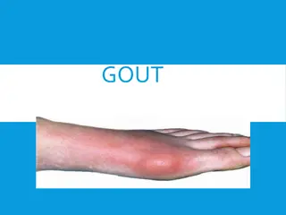
Gout Gout is one of the most metabolic painful rheumatic diseases. It results from deposits of needle-like crystals of uric acid in connective tissue, in the joint space between two bones, or in both. These deposits lead to inflammatory arthritis, which causes swelling, redness, heat, pain, and stiffness in the joints.
Gout ***Excess uric acid builds up in the joints and forms needle-like crystals that cause extreme pain. A gout attack can also be caused by joint injury, infection, kidney problems, medications or diet habits (e.g., a purine-rich, sugary diet, drinking too much alcohol or not enough water).
Pathophysiology Gout is a type of inflammatory arthritis (sometimes called gouty arthritis) that develops in people or animals who have high levels of uric acid in their body. Uric acid is produced naturally by the body and from eating foods that have purines. Purines are substances in animal and plant foods that the body converts to uric acid. Normally, the kidneys remove this acid from the blood and it leaves the body through the urine. With gout, excess uric acid builds up in the joints and forms needle-like crystals that cause extreme pain
CAUSES A gout attack can also be caused by joint injury, infection, kidney problems, medications or diet habits (e.g., a purine-rich, sugary diet, like meat, drinking too much alcohol or not enough water). Being overweight, having a family history of gout or having untreated high blood pressure, diabetes or metabolic syndrome also increases the risk of developing gout
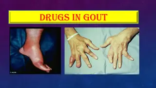
Gout Signs and Symptoms Gout attacks usually come on suddenly. animals may go to bed feeling fine but wake up with extreme joint pain. The first gout attack usually occurs in the large joint of the big toe. However, other joints and areas around the joints can be affected, like foot arches (insteps), ankles , heels and knees . Common symptoms include swelling, stiffness warmth and redness in and around joints. The pain may last hours or weeks. The build-up of uric acid can look and feel like lumps under the skin (tophi). It can also collect in the kidneys and cause small, hard deposits (kidney stones). , tenderness ,
Classification of gout Gout is classified as either articular (joint) gout or visceral (internal organ) gout. Gout usually occurs as one of two forms, visceral or articular. In visceral gout, the uric acid precipitates on internal organs, whereas articular gout occurs when uric acid tophi occur within and around joints and tendons.
Signs Visceral gout At post-mortem examination, chalky white urate deposits are found on all visceral surfaces and in renal tubules. Visceral gout is usually considered to be secondary to dehydration.
Articular urate deposition is less common and occurs after longterm increases in serum levels of uric acid. Deposits membranes in the toes and wing joints and incite a chronic granulomatous crystals (tophi). Joints are enlarged, and the feet appear deformed. Unlike visceral urate deposition, the kidneys are usually grossly normal. Articular urate deposition may be seen in birds that have hereditary defects in uric acid metabolism or that are fed excessive protein. develop on synovial reaction to urate
Microscopically, urate crystals are acicular, and they, or (more accurately) the spaces left after they have been dissolved by histologic processing, are surrounded macrophages, and multinucleated giant cells. by neutrophils,