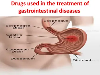
Gastro-Oesophageal Reflux Disease and Diagnostic Overview
Gastro-Oesophageal Reflux Disease (GORD) affects the upper gastrointestinal tract, causing symptoms like burning pain, regurgitation, and more. This condition is commonly associated with factors like Hiatus Hernia and Barrett's Oesophagus. Understanding the symptoms, diagnosis, and treatment options for GORD is essential for managing this prevalent condition effectively.

Download Presentation

Please find below an Image/Link to download the presentation.
The content on the website is provided AS IS for your information and personal use only. It may not be sold, licensed, or shared on other websites without obtaining consent from the author. If you encounter any issues during the download, it is possible that the publisher has removed the file from their server.
You are allowed to download the files provided on this website for personal or commercial use, subject to the condition that they are used lawfully. All files are the property of their respective owners.
The content on the website is provided AS IS for your information and personal use only. It may not be sold, licensed, or shared on other websites without obtaining consent from the author.
E N D
Presentation Transcript
Oesophagus Lecture II Dr. Zaeem Fezea Dahla Consultant General Surgeon CABS, MD
LEARNING OBJECTIVES Gastro-oesophageal disease (GORD) Barrett Oesophagus Hiatus Hernia Mallory -Weiss Syndrom Corrosive injuries
GASTRO-OESOPHAGEAL REFLUX DISEASE (GORD) The classical triad of symptoms is restrosternal burning pain Aetiology GORD is the most common condition affecting the upper gastrointestinal tract. Normal competence of the gastro-oesophageal junction is maintained by the LOS. This is influenced by both its physiological function and its anatomical location relative to the diaphragm and the oesophageal hiatus and cruri.
In GORD, LOS pressure tends to be generally low due to loss of an adequate length of intra-abdominal oesophagus (Hiatus Hernia). Crural opening widens allowing the upper stomach to slide up through the hiatus. The loss of the normal anatomical configuration exacerbates reflux. Sliding hiatus hernia is associated with GORD and may make it worse. Many GORD sufferers do not have a hernia, and many of those with a hernia do not have GORD
-The classical triad of symptoms is retrosternal burning pain(heart burn), epigastric pain (sometimes radiating through to the back) and regurgitation. -Most patients do not experience all three. -Symptoms are often provoked by food, particularly those that delay gastric emptying (e.g. fats, spicy foods).
As the condition becomes more severe, gastric juice may reflux to the mouth and produce an unpleasant taste often described as acid or bitter'. Heartburn and regurgitation can be brought on by stooping or exercise. A proportion of patients have odynophagia with hot beverages, citrus drinks or alcohol. Patients with nocturnal reflux and those who reflux food to the mouth nearly always have severe GORD
Diagnosis In most cases, the diagnosis is assumed rather than proven, and treatment is empirical. Investigation is only required when the diagnosis is in doubt, when the patient does not respond to a proton pump inhibitor (PPI) or if dysphagia is present. The most appropriate examination is endoscopy with biopsy.
In patients with atypical or persistent symptoms despite therapy, oesophageal manometry and 24- hour oesophageal pH recording (ideally with impedance measurement) may be justified to establish the diagnosis and guide management. 24-hour pH recording is the gold standard for diagnosis of GORD
. The endoscopic appearance of the normal squamous mucosa in the BODY OF ESOPHAGOUSER
The squamocolumnar junction is clearly seen in the lower oesophagus with a normal sharp demarcation.
Sliding hiatus hernia.The diaphragm can be seen constricting the upper stomach.
Management of uncomplicated GORD Medical management Most sufferers from GORD do not consult a doctor and do not need to do so. They self-medicate with over-the-counter medicines such as simple antacids, H2 blockers, or PPI. Simple measures that are often neglected include advice about weight loss, smoking, excessive consumption of alcohol, tea or coffee, the avoidance of large meals late at night and a modest degree of head-up tilt of the bed.
PPIs are the most effective drug treatment for GORD and patients are very reluctant to stop taking them once started on them. Given an adequate dose for 8 weeks, most patients have a rapid improvement in symptoms (within a few days), and more than 90 per cent can expect full mucosal healing at the end of this time. the dose of PPI is reduced after 8 weeks to that which keeps the patient free of symptoms, and this might even mean the cessation of PPI treatment.
Because most patients do not make major lifestyle changes and because PPIs are so effective, many remain on long-term treatment. For the minority who do not respond adequately to a standard dose, a trial at an increased dose or the addition of an H2-receptor antagonist is recommended. If unsuccessful, these patients should be formally investigated.
Surgery the number of antireflux operations has remained relatively constant and may even be increasing.This is probably due partly to increased patient expectations and partly to the advent of minimal access surgery, which has improved the acceptability of procedures. The indication for surgery in uncomplicated GORD is essentially patient choice. The risks and possible benefits need to be discussed in detail. Risks include a failed operation (5 10 per cent) and side effects such as dysphagia, gas bloat or abdominal discomfort (10 per cent).
With current operative techniques, 8590 per cent of patients should be satisfied with the result of an antireflux operation. Patients who are asymptomatic on a PPI need a careful discussion of the risk side of the equation. Reasons for failure on a PPI include: Volume reflux. Hermit lifestyle in which the least deviation from lifestyle rules leads to symptoms.
Psychological distress with intolerance of minor symptoms (a poor indication; these patients are likely to be dissatisfied with surgery) Poor compliance (a good indication if the reason for poor compliance is the side effects of treatment, otherwise a bad indication)
Which operation? There are many operations for GORD, but they are virtually all based on the creation of an intra-abdominal segment of oesophagus, crural repair and some form of wrap of the upper stomach (fundoplication) around the intra- abdominal oesophagus. (a) Total (Nissen) fundoplication; (b) partial fundoplication (Toupet).
Barrett's oesophagus (columnar-lined lower oesophagus) Barrett's oesophagus is a metaplastic change in the lining mucosa of the oesophagus in response to chronic gastro-oesophageal reflux Many of these patients do not have particularly severe symptoms, although they do have the most abnormal pH profiles. One of the great mysteries of GORD is why some people develop oesophagitis and others develop Barrett's oesophagus, often without significant oesophagitis.
Barrett's oesophagus with proximal migration of the squamocolumnar junction (a) and with a view of the distal oesophagus (b).
Barrett's oesophagus would need regular surveillance endoscopy with multiple biopsies in the hope of finding dysplasia or in situ cancer rather than allowing invasive cancer to develop and cause symptoms. Annual endoscopy has been widely practised, but two-year intervals are probably adequate, provided no dysplasia has been detected. Barrett's oesophagus was not diagnosed until there was at least 3 cm of columnar epithelium in the distal oesophagus. With the better appreciation of the importance of intestinal metaplasia, Barrett's oesophagus may be diagnosed if there is any intestinal metaplasia in the oesophagus.
Treatment of Barretts oesophagus When Barrett's oesophagus is discovered, the treatment is that of the underlying GORD. There has been considerable interest in recent years in endoscopic methods of ablating Barrett's mucosa in the hope of eliminating the risk of cancer development. Laser, photodynamic therapy, argon-beam plasma coagulation and endoscopic mucosal resection (EMR) have all been used
PARAOESOPHAGEAL (ROLLING) HIATUS HERNIA True paraoesophageal hernias in which the cardia remains in its normal anatomical position are rare. The vast majority of rolling hernias are mixed hernias in which the cardia is displaced into the chest and the greater curve of the stomach rolls into the mediastinum
paraoesophageal hernia showing the gastro-oesophageal junction just above the diaphragm and the fundus alongside the oesophagus, compressing the lumen. a
Potentially dangerous, because of volvulus The symptoms of rolling hernia are mostly due to twisting and distortion of the oesophagus and stomach. Dysphagia is common. Chest pain may occur from distension of an obstructed stomach. Classically, the pain is relieved by a loud belch. Symptoms of GORD are variable. Strangulation, gastric perforation and gangrene can occur.
Diagnosis of Paraosophageal hernia The hernia may be visible on a plain radiograph of the chest as a gas bubble, often with a fluid level behind the heart . A barium meal is the best method of diagnosis. The endoscopic appearances may be confusing, especially in large hernias when it is easy to become disorientated.
Treatment Symptomatic rolling hernias nearly always require surgical repair as they are potentially dangerous. Patients who present as an emergency with acute chest pain may be treated initially by nasogastric tube to relieve the distension that causes the pain, followed by operative repair. If the pain is not relieved or perforation is suspected, immediate operation is mandatory.
A gas bubble seen on a plain chest radiograph, showing the fundus of the stomach in the chest.
MALLORY-WEISS SYNDROME Forceful vomiting may produce a mucosal tear at the cardia rather than a full perforation. The mechanism of injury is different. In Boerhaave's syndrome, vomiting occurs against a closed glottis, and pressure builds up in the oesophagus. In Mallory Weiss syndrome, vigorous vomiting produces a vertical split in the gastric mucosa, immediately below the squamocolumnar junction at the cardia in 90 per cent of cases. The condition presents with haematemesis. Usually, the bleeding is not severe, but endoscopic injection therapy may be required for the occasional, severe case.
The endoscopic appearance of a mucosal tear at the cardia.
CORROSIVE INJURY Corrosives such as sodium hydroxide or sulphuric acid may be taken in attempted suicide. Accidental ingestion occurs in children and when corrosives are stored in bottles labelled as beverages. All can cause severe damage to the mouth, pharynx, larynx, oesophagus and stomach. The type of agent, its concentration and the volume ingested largely determine the extent of damage. In general, alkalis are relatively odourless and tasteless, making them more likely to be ingested in large volume.
Alkalis cause liquefaction, saponification of fats, dehydration and thrombosis of blood vessels that usually leads to fibrous scarring. Acids cause coagulative necrosis with eschar formation, and this coagulant may limit penetration to deeper layers of the oesophageal wall. Acids also cause more gastric damage than alkalis because of the induction of intense pylorospasm with pooling in the antrum. Symptoms and signs are notoriously unreliable in predicting the severity of injury.
The key to management is early endoscopy by an experienced endoscopist to inspect the whole of the oesophagus and stomach These patients can safely be fed.With more severe injuries, a feeding jejunostomy may be appropriate until the patient can swallow saliva satisfactorily. The widespread use of broad-spectrum antibiotics and steroids is not supported by evidence.
Caustic or lye stricture with marked stenosis high in the body of the oesophagus.The strictures are frequently multiple and difficult to dilate unless treated energetically at an early stage.
THANK YOU


")


 HIATUS")