Gastroesophageal reflux disease (GERD) is a common condition where gastric juice refluxes into the esophagus, leading to symptoms and potential mucosal injury. This article covers the pathophysiology of GERD, distinguishing between physiologic and pathologic reflux, the role of the lower esophageal sphincter, and factors contributing to abnormal reflux. Understanding GERD can help in recognizing clinical features, complications, and management of this condition.
Please find below an Image/Link to download the presentation.
The content on the website is provided AS IS for your information and personal use only. It may not be sold, licensed, or shared on other websites without obtaining consent from the author. If you encounter any issues during the download, it is possible that the publisher has removed the file from their server.
You are allowed to download the files provided on this website for personal or commercial use, subject to the condition that they are used lawfully. All files are the property of their respective owners.
The content on the website is provided AS IS for your information and personal use only. It may not be sold, licensed, or shared on other websites without obtaining consent from the author.
E N D
Presentation Transcript
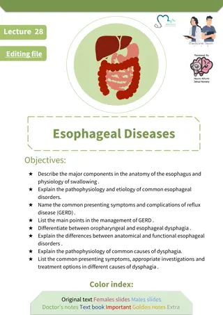
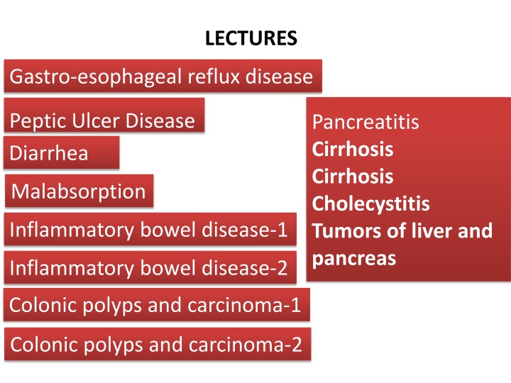
LECTURES Gastro-esophageal reflux disease Peptic Ulcer Disease Diarrhea Pancreatitis Cirrhosis Cirrhosis Cholecystitis Tumors of liver and pancreas Malabsorption Inflammatory bowel disease-1 Inflammatory bowel disease-2 Colonic polyps and carcinoma-1 Colonic polyps and carcinoma-2
Objectives Upon completion of this lecture the students will : 1. Define gastroesophageal reflux disease 2. Understand the Pathophysiology of reflux esophagitis. 3. Know clinical features of reflux esophagitis 4. Describe the pathological features of reflux esophagitis 5. Know the complications of reflux esophagitis
Gastroesophageal Reflux Disease (GERD) Gastroesophageal reflux is a normal physiologic phenomenon experienced intermittently by most people, particularly after a meal. Gastroesophageal reflux disease (GERD) occurs when the amount of gastric juice that refluxes into the esophagus exceeds the normal limit, causing symptoms with or without associated esophageal mucosal injury.
Physiologic vs Pathologic Physiologic GERD Postprandial Short lived Asymptomatic No nocturnal symptomes Pathologic GERD Symptoms Mucosal injury Nocturnal symptomes
GERD Pathophysiology Abnormal lower esophageal sphincter or Increase abdominal pressure
GERD Pathophysiology A. Abnormal lower esophageal sphincter 1. Functional (frequent transient LES relaxation) 2. Mechanical (hypotensive LES) 3. Foods (eg, coffee, alcohol), 4. Medications (eg, calcium channel blockers), 5. Location .......... hiatal hernia (x ray show gas behind the heart) The most common cause of (GERD). decrease the pressure of the LES. or obesity Pregnancy increased gastric volume B. Increase abdominal pressure
Pathophysiology Primary barrier to gastroesophageal reflux is the lower esophageal sphincter LES normally works in conjunction with the diaphragm If barrier disrupted, acid goes from stomach to esophagus
Clinical Manisfestations 85638LRtQ_w Most common symptoms Heartburn retrosternal burning discomfort Regurgitation effortless return of gastric contents into the pharynx without nausea Atypical symptoms .coughing, chest pain, and wheezing.
Diagnostic Evaluation If classic symptoms of heartburn and regurgitation exist in the absence of alarm symptoms the diagnosis of GERD can be made clinically and treatment can be initiated
Esophagogastrodudenoscopy Endoscopy (with biopsy if needed) In patients with alarm signs/symptoms Those who fail a medication trial
Complications Erosive esophagitis Stricture Barrett s esophagus
Complications Erosive esophagitis Responsible for 40-60% of GERD symptoms Severity of symptoms often fail to match severity of erosive esophagitis
Esophagitis Eosinophils and neutrophils Elongation of lamina propria papillae basal zone hyperplasia,
Complications Esophageal stricture Result of healing of erosive esophagitis May need dilation
Complications 8-15% Barrett s Esophagus Intestinal metaplasia of the esophagus Associated with the development of adenocarcinoma
Complications Barrett s Esophagus Acid damages lining of esophagus and causes esophagitis Damaged area heals in a metaplastic process and abnormal columnar cells replace squamous cells This specialized intestinal metaplasia can progress to dysplasia and adenocarcinoma Many patients with Barrett s are asymptomatic
Treatment H 2 receptor Blockers Proton pump inhibitors Antireflux surgery
Objectives Define gastroesophageal reflux disease Symptoms OR mucosal damage produced by the abnormal reflux of gastric contents into the esophagus Physiologic vs. pathologic Understand the Pathophysiology of reflux esophagitis. Abnormal lower esophageal sphincter or increased abdominal pressure Know clinical features of reflux esophagitis Heartburn, Regurgitation Atypical symptoms .coughing, chest pain, and wheezing Describe the pathological features of reflux esophagitis Eosinophils and neutrophils Elongation of lamina propria papillae basal zone hyperplasia Erosive esophagitis, Stricture, Barrett s esophagus ..dysplasia .adenocarcinoma Know the complications of reflux esophagitis
Objectives Upon completion of this lecture the students will : 1. Understand the Pathophysiology of acute and chronic peptic ulcer 2. Know the possible causes of gastric and duodenal ulcers with emphasis on most common causes (H pylori and drugs) 3. Recognize the gross and microscopic features of peptic ulcer 4. Recognize the clinical features and consequences of acute and chronic peptic ulcer
Ulcer Ulcer: a breach in the mucosa of the alimentary tract extending through muscularis mucosa into submucosa or deeper. 1. Peptic ulcer 2. Stress ulcers (acute gastric ulcers)
Acute peptic ulcers Pathophysiology As part of an acute gastritis As a complication of a severe stress response As a result of extreme hyperacidity.
Pathophysiology Acute peptic ulcers As part of an acute gastritis (acute response to an irritant 'chemical' injury by drugs (NSAID) or alcohol As a complication of a severe stress response (severe burns (Curling's ulcer), major trauma (cushing ulcer) or cerebrovascular accidents ) As a result of extreme hyperacidity (Zollinger- Ellison syndrome).
Chronic peptic ulcer Peptic Ulcer Disease Locations May occur in any portion of the GI tract exposed to acidic gastric juices 98% located in first portion of duodenum or stomach, ratio = 4:1 Esophagus . as a result of GERD or acid secretion by ectopic gastric mucosa. Gastric mucosa within a Meckel diverticulum can result in peptic ulceration of adjacent mucosa.
Peptic Ulcer Disease Gastric ulcers Pathophysiology The mucosal defences against acid attack consist of: 1. Mucus-bicarbonate barrier 2. The surface epithelium.
Peptic Ulcer Disease Gastric ulcers Pathophysiology The mucosal defences against acid attack consist of: 1. Mucus-bicarbonate barrier Duodeno-gastric reflux ( bile ) 2. The surface epithelium. 1. NSAIDs (blocking the synthesis of the prostaglandins) 2. H. pylori infection, ( cytotoxins and ammonia) Thus peptic ulcers in the stomach, breakdown of mucosal defence is much more important than excessive acid production.
Peptic Ulcer Disease Duodenal ulcers Pathophysiology Increased production of acid assumes more importance in the pathogenesis of duodenal ulceration H. pylori-infected individuals secrete 2-6 times as much acid as non-infected controls Helicobacter Pylori does not colonise normal duodenal epithelium Helicobacter is involved in duodenal ulceration because there is gastric metaplasia in response to excess acid. Gastric metaplasia paves the way for colonisation by Helicobacter + =Duodenal ulcers Increased production of acid Helicobacter P
Peptic Ulcer Disease Pathophysiology Duodeno-gastric reflux ( bile ) Hyperacidity Gastric ulcers Duodenal ulcers H. pylori NSAIDs H pylori infection of the pyloric antrum is present in nearly all patients with chronic duodenal ulcer and approximately 75% of patients with chronic gastric ulcer. Although more than 70% of individuals with PUD are infected by H. pylori, fewer than 20% of H. pylori infected individuals develop peptic ulcer.
Morphology Gross usually less than 20 mm in diameter but they may > 100 mm in diameter. The classic peptic ulcer is a round to oval, sharply punched-out defect In contrast, heaped-up margins are more characteristic of cancers Microscopy the base consists of necrotic tissue and polymorph exudate overlying inflamed granulation tissue which merges with mature fibrous (scar) tissue.
Clinical features Epigastric pain (the most common symptom) Gnawing or burning sensation Occurs 2-3 hours after meals Relieved by food or antacids Patient awakens with pain at night. Some present with complications such as iron deficiency anemia, frank hemorrhage, or perforation.
Therapy Current therapies for PUD are aimed at I. II. Acid suppression H. pylori eradication a) Proton pump inhibitors b) H2 blockers
Objectives Understand the Pathophysiology and etiology of acute and chronic peptic ulcer Understand the Pathophysiology and etiology of acute and chronic peptic ulcer Increase Aggressive Factors or dercrease Defensive Factors Acute ulcers ..acute gastritis , severe stress response, extreme hyperacidity Gastric: H. pylori infection, NSAIDs , bile reflux Chronic ulcer Duodenal: H. pylori infection , hyperacidity Recognize the gross and microscopic features of peptic ulcer Recognize the gross and microscopic features of peptic ulcer The classic peptic ulcer is a round to oval, sharply punched-out defect necrotic tissue and polymorph exudate overlying inflamed granulation tissue which merges with mature fibrous (scar) tissue. Recognize the clinical features and consequences of acute and chronic peptic ulcer Recognize the clinical features and consequences of acute and chronic peptic ulcer Epigastric pain Gnawing or burning sensation Some present with complications such as iron deficiency anemia, frank hemorrhage, or perforation


")