General Histopathology EQA Scheme Meeting Agenda and Review
Join the South East England General Histopathology EQA Scheme for a Case Discussion Round where participants will review cases, discuss meeting etiquette, and adhere to important meeting terms of reference. This educational exercise offers insights into scoring, merging decisions, and diagnostic categories. A must-attend event for histopathology professionals on Wednesday, 27th November, 2024
Uploaded on | 3 Views
Download Presentation

Please find below an Image/Link to download the presentation.
The content on the website is provided AS IS for your information and personal use only. It may not be sold, licensed, or shared on other websites without obtaining consent from the author. If you encounter any issues during the download, it is possible that the publisher has removed the file from their server.
You are allowed to download the files provided on this website for personal or commercial use, subject to the condition that they are used lawfully. All files are the property of their respective owners.
The content on the website is provided AS IS for your information and personal use only. It may not be sold, licensed, or shared on other websites without obtaining consent from the author.
E N D
Presentation Transcript
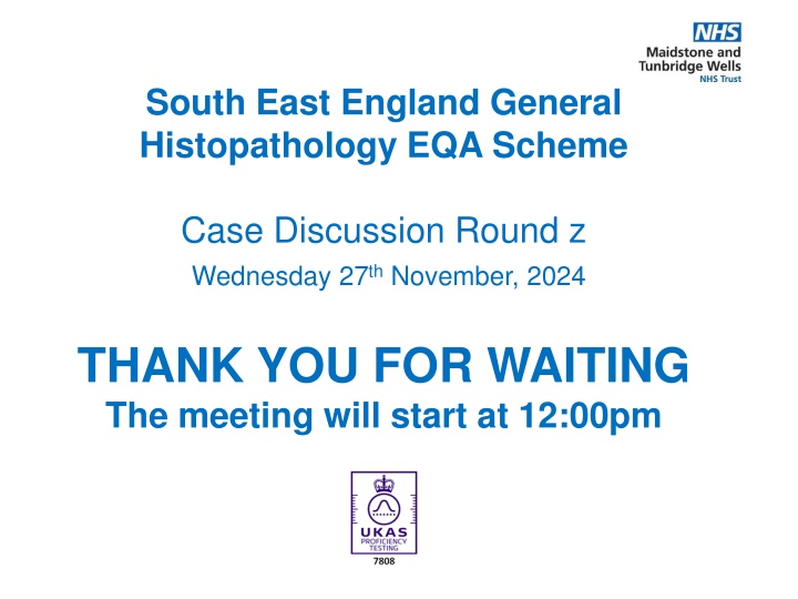
South East England General Histopathology EQA Scheme Case Discussion Round z Wednesday 27thNovember, 2024 THANK YOU FOR WAITING The meeting will start at 12:00pm
Meeting Etiquette 4 3 2 1 Mute your mic if you re not speaking Wait for the Chair person to call on you before you unmute your mic Use the raise hand Or chat feature to raise questions or share ideas If your camera is on, everyone can see you Remember Everyone can see your chat comments 6
Agenda 1. Welcome & Introduction of Scheme Staff 2. Meeting Terms of Reference 3. Case and Preliminary Score Review a) Case 939-948 b) Educational Cases 949-950 4. Questions / comments
This meeting is held between the end of case consultation and results being issued and now replaces the additional final week of the case consultation. This meeting is an educational exercise; an opportunity to explain the reasons behind scoring and merging or why cases were excluded. For clarity, this is not an opportunity to alter merging decisions, as participants have that opportunity during the Case Consultation period. An additional CPD point will be awarded to those who attend, and it will be added to the annual certificate. Please note you have to stay for >50% of the meeting to gain this point (attendance times are monitored automatically by Teams) We always welcome any feedback good or bad you may have about today.
CaseConsultation 163 responses received for round z 87 responses received for consultation 53.3% QUORATE Thank-you for submitting responses and consultation on time you have made completion of this round much easier for all Basic Rules regarding Case Consultation and Merging Diagnostic categories: If you are exempt from a category, your consultation response to that case is not counted Each case must have received a consultation response from at least 50% of those that answered it For a merge to be automatically accepted, more than 50% of consultation respondents must agree Between 40-50% agreement, the merge will be accepted only with the agreement of the Organiser (i.e. clinically valid). The consensus CAN be over-ridden if there are clinically valid reasons for doing so. These are recorded, and reviewed at the AMR.
Case 939 GU Specimen: Penile Lesion Submitted Diagnosis: Well differentiated squamous cell carcinoma Clinical Macro Immuno Image link Preliminary Results Final Merge Results Submitted M48. Penile lesion, proliferative Multiple pieces of soft brown tissue, together in 70x40x20mm P40 positive, P16 negative Click here to view digital image 1. SCC. HPV Not mentioned 4.84 2. SCC. HPV Independent 2.95 3. SCC. HPV Dependent 0.13 4. No invasion (PeIN / SIL 0.26 / severe squamous dysplasia) 5. Verruciform / Verrucous carcinoma 1.12 6. PeIN / SIL / sev sq dysplasia - invasion possible. 0.49 Non HPV 7. Condyloma Accuminatum 0.21 1, 2, 3, 5 Most participants agreed to merge 1, 2, 3 however 5 will also be merged as this is also clinically valid
Case 940 Endocrine Specimen: Thyroid Submitted Diagnosis: Papillary Thyroid carcinoma. Clinical Macro Immuno Image link Preliminary Results Final Merge Results M64.Multiple pathological left lateral cervical lymph nodes Thyroid gland weighing in total 28g. Left lobe measures 39x16x20mm, right lobe 45x25x35mm and isthmus 15x14x5mm. None Provided Click here to view digital image 1. Papillary Carcinoma 7.42 2. Papillary Carcinoma - Follicular Variant 1.96 3. Papillary Carcinoma - Tall cell variant 0.50 4. Papillary Carcinoma - Columnar Cell Type 0.06 5. Papillary Carcinoma - Infiltrative 0.06 follicular type 56.3% of participants agreed to merge all diagnostic categories On slicing the left lobe, there is a solid, white lesion measuring 22mm in diameter that is close to the specimen margin. The remaining thyroid tissue appears unremarkable.
Case 941 Respiratory Specimen: Core Lung Biopsy Submitted Diagnosis: Recurrent Aspergilloma Clinical Macro Immuno Image link Preliminary Results Final Merge Results F59, core biopsy lung. Previous aspergilloma. Right upper lobe opacity (stable) 67.5% of participants agreed to merge 1 and 3 Several cores 5- 10mm PAS+ / Grocott +ve Click here to view digital image 1. Aspergillosis 9.17 2. Pneumocystis 0.12 3. Fungal infection 0.71
Case 942 Lymphoreticular Specimen: Bone Marrow Trephine Submitted Diagnosis: Leishmaniasis Clinical Macro Immuno Image link Preliminary Results Final Merge Results 41% participants agreed to no merging on this case. M61. Cytopenia, myeloma.? Progression on treatment. Haemorrha gic core 22mm. CD138 positive cells present (1- 2%), do not express CD56, cyclin D1 or CD20. Click here to view digital image 1. Leishmaniasis 8.47 2. Histoplasmosis 0.75 3. Parasitic infection 0.25 4. Blastomycosis 0.02 5. Aspergillosis 0.07 6. Smoldering multiple myeloma 0.07 7. Toxoplasmosis 0.01 8. Fungal infection 0.15 9. No evidence of progression. Discuss 0.14 with Haematopathology colleagues 10. Increased Haematopoeisis. Refer to 0.07 lymphoma panel No Light chain restriction.
Case 943 Gynae Specimen: Polyp Endometrial curetting s Submitted Diagnosis: Low grade endometrial stromal tumour (differential diagnosis includes endometrial stromal sarcoma and endometrial stromal nodule) Clinical Macro Immuno Image link Preliminary Results Final Merge Results Diagnoses 1, 2 ,3, 4 & 5 will be merged. F57. Perimenopausal bleeding. TVS scan shows 8mm ET and submucous fibroids with polyps. Bulky tan and haemorrhagic fragments 40mm in aggregate. Positive: CD10, SMA, Desmin, WT1, ER and PR. Click here to view digital image 1. Endometrial Stromal sarcoma - high grade 0.00 2. Endometrial Stromal sarcoma - low grade 6.13 3. Endometrial Stromal sarcoma - grade not 1.95 mentioned 4. Stromal Nodule 0.97 5. Stromal neoplasm / tumour 0.76 6. Stromal Poly / Stromal polyp 0.06 7. Cellular leiomyoma 0.10 8. Stromo-myoma 0.03 Negative: Cytokeratin (A1/3), CD117 and CD34. 52% of participants agreed to merge 1,2&3 however 4 and 5 have been included as clinical details to not allow precise distinction Wild type p53 staining. Diagnostic hysteroscopy done and multiple polyps removed (friable) and curette is sent.
Case 944 GI Specimen: Core of liver Submitted Diagnosis: Angiomyolipoma Clinical Macro Immuno Image link Preliminary Results Final Merge Results F70. Evidence of segment 2 HCC currently measuring 19mm. One core of liver 13mm and 1 mm in diameter. Negative: CK7, CK19, CK20, CAM 5.2, AE1/3, CD10, glypican 3 and CD68. : Positive: HMB45, MelanA, SMA, calponin and weak CD117. Click here to view digital image 1. Angiomyolipoma / PEComa 9.44 2. Melanoma 0.19 3. Clear cell sarcoma 0.06 4. Clear cell sugar (epithelial) tumour 0.12 5. Metastatic malignant perivascular 0.12 epithelioid tumour 6. PECOMA mets 0.06 7. neoplasm of uncertain malignant potential 0.01 - myomelanocytic differentiation 46% participants agreed to no merging on this case
Case 945 Skin Specimen: Skin Submitted Diagnosis: Regressing keratoacanthoma Clinical Macro Immuno Image link Preliminary Results Final Merge Results M88. Cyst of sternum EOS 11 x 10mm with irregular raised lesion 10 x 5mm. None Provided Click here to view digital image 1. Keratocathoma 4.85 2. SCC (karatocanthoma-like well differentiated) 1.11 3. Hyperplasia / epidermal naevus / Acantholytic 0.44 acanthoma 4. Squamoproliferative lesion / benign papillary 1.42 lesion 5. Seborrhoeic keratosis / inverted follicular 0.60 keratosis 6. Viral wart / dermatophyte / granulomatous 0.88 inflammation / fungal 7. Warty Dyskeratoma 0.06 8. Lichenoid actinic keratosis 0.42 9. Atypical Squamoproliferative lesion 0.06 10. Ruptured epidermal cyst with inflammation 0.16 This case will be excluded from personal scores. Even if the 3 most popular diagnoses were merged, we would not be able to achieve consensus
Case 946 Breast Specimen: Breast Submitted Diagnosis: Myofibroblastoma Clinical Macro Immuno Image link Preliminary Results Final Merge Results F70. B3 lump right breast. 12g of breast tissue containing a circumscribed grey tumour (17m in diameter) Immuno (in previous biopsy) Positive for CD34, bcl-2, ER, PR. Negative for S100, CK7, MNF116 and SMA. Click here to view digital image 1. Angiolipoma 0.14 2. Myofibroblastoma 7.79 3. Angiofibroma 0.42 4. Haemangiopericytoma / solitary 0.80 fibrous tumour 5. Angiomyofibroblastoma 0.37 6. Haemangioma 0.26 7. Hamartoma with myofibroblastic 0.13 component 8. Benign spindle cell lesion 0.03 9. Pleomorphic hyalinizing angiectatic 0.06 tumour Diagnoses 2 and 5 will be merged
Case 947 Miscellaneous Specimen: Mass from finger Submitted Diagnosis: Ancient Schwannoma Clinical Macro Immuno Image link Preliminary Results Final Merge Results M61. Lump left little finger? GCT 64g. 55 x 50 x 40 mm. Smooth surface. None Provided Click here to view digital image 1. Schwannoma / Neurilemmoma 7.84 2. Rheumatoid nodule 0.28 3. Giant Cell Tumour / fibroma of 0.25 Tendon Sheath / glomus tumour 4. Vascular lesions incl haemangioma 0.56 / AV malformation etc 5. Epithelioid sarcoma 0.18 6. Massons tumour 0.51 7. Neurofibroma 0.06 8. Organising Thrombus / granuloma 0.13 annulare / gout 9. Angiosarcoma 0.03 10. Pleomorphic hyalinizing angiectatic 0.16 tumour /bone cyst No merges (73.56% of participants)
Case 948 Digital Only - Miscellaneous Specimen: Bone Submitted Diagnosis: Metastatic carcinoma, favouring metastatic salivary duct carcinoma. (breast carcinoma was P53 and Her 2 strongly positive). Clinical Macro Immuno Image link Preliminary Results Final Merge Results : F73. Bone metastases - biopsied. PET: positive 9mm breast lesion (grade 3 carcinoma with apocrine differentiation Her-2 positive). 2 pieces of tissue, the larger measuring 5 x 2mm and the smaller 4 x 2 mm. Tumour positive for AE1/AE3, GATA3 and androgen receptor. PSA, P53 and Her-2 negative. Click here to view digital image 1. Metastatic Salivary Duct Carcinoma 3.74 2. Metastatic Breast carcinoma 2.91 3. Metastatic carcinoma - clinical correlation 0.67 required 4. Metastatic carcinoma - compare with previous 0.06 (breast or salivary) 5. Metastatic carcinoma (acinic cell) 0.11 6. Metastatic carcinoma - NOS or breast/salivary 2.38 7. Metastatic apocrine carcinoma 0.12 8. TCC 0.01 This case will be excluded from personal scoring due to lack of consensus Previous history of left buccal salivary duct carcinoma (pT2 pN0 with perineural invasion)
Case 949 EDUCATIONAL Specimen: Maxilla Biopsy Clinical Macro Immuno Image link Suggested Diagnosis (Top 5) Submitted Diagnosis M34. Soft tissue tumour between UL2 and UL3 anterior maxilla. Punch biopsy 6mm x 5mm depth AE1/3 and Conga Red Positive S100, CD68 and TTF1 Negative Proliferation fraction very low. . Click here to view digital image 1. Calcifying epithelial odontogenic tumour x 52 2. Pindborg tumour x 8 3. Calcifying odontogenic tumour x 8 4. Calcifying epithelial odontogenic x 6 (Pindborg) tumour 5. Amyloid producing odontogenic tumour x 6 Calcifying Epithelial Odontogenic Tumour.
Case 950 EDUCATIONAL Specimen: Skin Excision Clinical Macro Immuno Image link Suggested Diagnosis (Top 5) Submitted Diagnosis M77. Scalp, 2- month history of fast-growing skin lesion, exophytic and ulcerated. Disc of oriented skin measuring 29 x 29 x 5 mm with an ulcerated nodule measuring 20mm in diameter. Positive for SMA, negative for AE1/AE3, Desmin, CD34, factor Xllla, P40 and SOX10 Click here to view digital image 1. Pleomorphic dermal sarcoma x 42 2. Atypical fibroxanthoma x 35 3. Leiomyosarcoma x 15 4. ATYPICAL FIBROXANTHOMA x 12 5. AFX x 7 Leiomyosarcoma
4. Questions Comments Suggestions Feedback Thank you for attending. This presentation can be found on the EQA website from next week.

