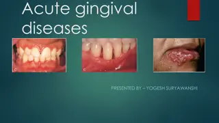
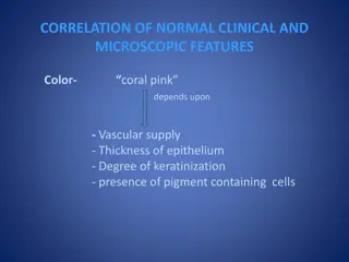
GINGIVAL INFLAMMATION
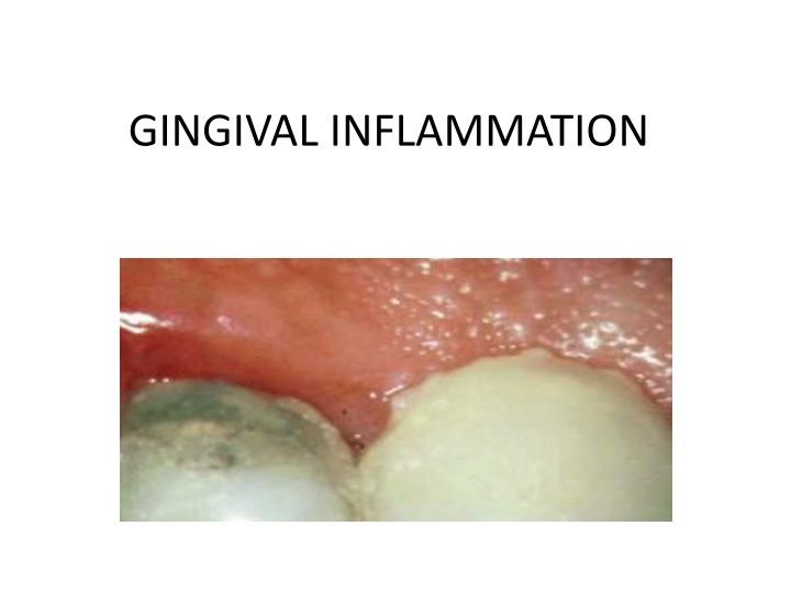
Gingival inflammation progresses through various stages, from initial vascular changes to established lesions. Microscopic examination reveals cellular infiltrates and collagen destruction. Learn about the evolution of gingivitis and its physiological impacts.

Download Presentation

Please find below an Image/Link to download the presentation.
The content on the website is provided AS IS for your information and personal use only. It may not be sold, licensed, or shared on other websites without obtaining consent from the author.If you encounter any issues during the download, it is possible that the publisher has removed the file from their server.
You are allowed to download the files provided on this website for personal or commercial use, subject to the condition that they are used lawfully. All files are the property of their respective owners.
The content on the website is provided AS IS for your information and personal use only. It may not be sold, licensed, or shared on other websites without obtaining consent from the author.
E N D
Presentation Transcript
INTRODUCTION Stage I Gingivitis(Initial lesion) Stage II Gingivitis(Early lesion) Stage III Gingivitis(Established lesion) Stage IV Gingivitis(Advanced lesion)
STAGE I GINGIVITIS(INITIAL LESION) First manifestation Vascular changes Dilated capillaries Blood flow increased Initial inflammatory changes due to microbial activation of resident leukocytes & subsequent stimulation of endothelial cells Subclinical gingivitis
STAGE I GINGIVITIS(INITIAL LESION) Contd Microscopically: -widening of small capillaries/venules -adherence of neutrophils to vessle walls (margination)-1wk/early as 2 days after plaque accumulation -PMNs(diapedesis,emigration) CT,JE&GS -Exudation of fluid from GS&extravascular protein are present
STAGE II GINGIVITIS(EARLY LESION) Evolves from the initial lesion After 1wk of plaque accumulation Erythema Bleeding on Probing Gingival fluid flow Transmigrating leukocyte maximum-6&12days
STAGE II GINGIVITIS(EARLY LESION)Contd Microscopic examination: -Leucocyte(mainly Lymphocyte-75%T Cells)infilterate in CT below JE -Some neutrophils,macrophages,plasma cells&mast cells -JE densely infilterated with neutrophils&show development of rete pegs/ridges
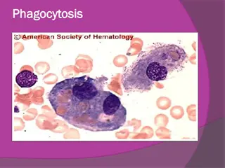
STAGE II GINGIVITIS(EARLY LESION)Contd. 70%collagen destruction around cellular infilterate Circular&Dentogingival group fibers mostly affected PMNs reach in to pocket(chemotactic stimuli) PMNs-phagocytosis(release of lysosomes to engulf bacteria) MMPs-extracellular matrix remodelling(7days)
STAGE III GINGIVITIS(ESTABLISHED LESION) 2/3wk after accumulation of plaque Predominance of plasma cells&B lymphocyte Creation of small gingival pocket lined with pocket epithelium B cells predominently IgG1&IgG3 subclasses Vessels engorged ,congested&venous return impaired&blood flow sluggish Result localized gingival anoxemia-bluish hue on the reddened gingiva
STAGE III GINGIVITIS(ESTABLISHED LESION contd.. Extravastion of CT&brekdown in to component pigments can deepen the color of inflamed gingiva Collagenolytic activity increased in inflammed gingival tissue Increased level glucuronidase,glactosidase,glucocidase,estera se,aminopeptidase&cytochrome oxidase Decreased neutral mucopolysaccharide erythrocyte in to of ALP, -
STAGE III GINGIVITIS(ESTABLISHED LESION contd.. Microscopically: -Increased number of Plasma cells -JE, widened cellular debries including lysosomes -JE develop ridges/rete peges protrudes in CT -Collagen fibers destroyed This lesion is reversible Periodontal therapy intercellular spaces+granular after successful
STAGE IV GINGIVITIS(ADVANCED LESION) Phase of periodontal breakdown Extension of lesion into alveolar bone Higher IL-1 &lower IL-8 at 28days Microscopic: -fibrosis of gingiva -widespread manifestation of inflammatory and immunologic tissue damage -plasma cell dominates in CT -neutrophils dominates JE
CONCLUSION Time (days) Blood vessels JE&SE Predomin ent cell Collagen C/F Initial lesion 2-4 vasculitis PMNs PMNs Perivascul ar loss GCF flow Early lesion 4-7 Vascular proliferati on Same as stage I rete pegs Lymphocy te Loss around infiltrate Erythema BOP Establishe d lesion 14-21 As stage II +Blood stasis As stage II +more advanced Plasma cells Continued loss Changes in color,size &texture etc
MCQ-1 Which represents the non apparent initial response of the gingiva to bacterial plaque (a)Subclinical gingivitis (b)Marginal gingivitis (c)Localized gingivitis (d)Generalized gingivitis of the following clinical condition
MCQ-2 Which of the following leucocytes mainly predominant in the stage I gingivitis (a)PMNs (b)Plasma cells (c)Lymphocytes (d)Basophils
MCQ-3 Which of the following is related with clinical finding of the early lesion of gingivitis (a)Gingival fluid flow (b)Erythema (c)Change texture of gingiva (d)Change size of gingiva
MCQ-4 Which of the following cell is mainly present in the C.T.during stage II gingivitis (a)Neutrophils (b)Lymphocyte (c)Plasma cell (d)Macrophages
MCQ-5 Which group of the fiber is mostly affected in the early stage of gingivitis (a)Circular (b)Alveolar crest (c)Horizontal (d)Transseptal
MCQ-6 The superimposes a some what bluish hue on the reddened gingiva is clinical feature of (a)Initial lesion (b)Early lesion (c)Established lesion (d)Advanced lesion localized gingival anoxemia which
REFERENCES Carranza s Clinical Periodontology 11thEdition Carranza s Clinical Periodontology 12thEdition


")
")
")