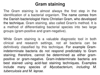
Gram Stain in Bacteriology

The Gram stain is a fundamental tool in microbiology for distinguishing between Gram-positive and Gram-negative bacteria based on cell wall characteristics. The stain involves multiple steps using crystal violet, iodine, ethanol, and safranin, resulting in bacteria appearing blue/purple or pink/red under a microscope. Gram-positive bacteria have a simpler cell wall structure with a thick peptidoglycan layer, while Gram-negative bacteria have a more complex cell wall with a thin peptidoglycan layer and an outer lipopolysaccharide layer. Explore the principles and differences between these bacterial types.

Download Presentation

Please find below an Image/Link to download the presentation.
The content on the website is provided AS IS for your information and personal use only. It may not be sold, licensed, or shared on other websites without obtaining consent from the author.If you encounter any issues during the download, it is possible that the publisher has removed the file from their server.
You are allowed to download the files provided on this website for personal or commercial use, subject to the condition that they are used lawfully. All files are the property of their respective owners.
The content on the website is provided AS IS for your information and personal use only. It may not be sold, licensed, or shared on other websites without obtaining consent from the author.
E N D
Presentation Transcript
Gram stain Dr. Mayssaa Essam
Principle The Gram stain is the most useful and widely employed differential stain in bacteriology. It divides bacteria in to two groups (Gram positive and Gram negative bacteria. The primary stain is crystal violet. It is followed by treatment with an iodine solution , which function as a mordant , that is , it increase the interaction between the bacterial cell and the dye .
The smear than decolorized by washing with an agent such as 95% ethanol. Gram positive bacteria retain the crystal violet-iodine complex when washed with the decolorizer where as gram negative bacteria lose their crystal violet-iodine complex and become colorless. Finally , the smear is counter stianed with a basic dye, different in color than crystal violet. This is safranin, the safranin will stain the colorless gram negative bacteria pink but does not alter the dark purple color of gram positive bacteria.
Difference between Gram positive and Gram negative bacteria Gram positive bacteria Simple cell wall Gram negative bacteria More complex cell wall Thick peptidoglycan cell wall layer Thin peptidoglycan cell wall layer No outer lipopolysaccharide wall layer Retain crystal violat/iodine outer lipopolysaccharide wall layer Retain safranin Appear (Blue/Purple) Appear (Pink/Red)
Staphylococcus aureus Escherichia coli