Guidelines for Developing Radiogenomic Research Teams in Cancer Studies
This resource outlines the processes for establishing successful radiogenomic research teams in cancer studies, based on the experiences of teams working with various tumor types like GBM, BRCA, KIRC, and OV. The guidelines cover aspects from data acquisition to team formation and governance, providing a roadmap for researchers in the field.

Download Presentation

Please find below an Image/Link to download the presentation.
The content on the website is provided AS IS for your information and personal use only. It may not be sold, licensed, or shared on other websites without obtaining consent from the author.If you encounter any issues during the download, it is possible that the publisher has removed the file from their server.
You are allowed to download the files provided on this website for personal or commercial use, subject to the condition that they are used lawfully. All files are the property of their respective owners.
The content on the website is provided AS IS for your information and personal use only. It may not be sold, licensed, or shared on other websites without obtaining consent from the author.
E N D
Presentation Transcript
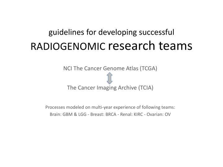
guidelines for developing successful RADIOGENOMIC research teams NCI The Cancer Genome Atlas (TCGA) The Cancer Imaging Archive (TCIA) Processes modeled on multi-year experience of following teams: Brain: GBM & LGG - Breast: BRCA - Renal: KIRC - Ovarian: OV
Starting framework Start expert team formation once pre-surgical tumor type TCIA image collection has reached statistically robust size of > 100 cases Log/list inventory of scan types of each case available (e.g. CT type, MR pulse sequences, etc) https://wiki.cancerimagingarchive.net/display/Public/Wiki List/publish clinical data field available from TCGA Data Portal for that tumor type https://tcga-data.nci.nih.gov/tcga/tcgaHome2.jsp
Next steps Contact interested clinical image experts Ask them to outreach to expert image reader colleagues known to them (~ a dozen or so) Ask or suggest advisory colleagues with tumor relevant expertise in genetics, oncology, stats Define governance/communication processes T-con schedule; reading parameters; collegial and publication/authorship/citation understandings
Data acquisition processes Team generates v.1 structured-reporting CRF feature set pick-list with quantitative elements Team constructs Visual Manual depicting each feature listed choice element NCI CIP creates Wiki and Google-doc sites specific to team Training set case-list created sparing TCGA cases if possible
Example TCGA Breast Imaging Scoring Visual Guide v8 Based on BI-RADS, but more granular
Example: Breast Feature List 1.1 Breast Observations Extent 1.2 Breast Observations - Flag for extensive heterogeneous disease 1.3 Breast Observations - Fibroglandular 1.4 Breast Observations Background 1.5 Breast Observations - Associated Features 1.6 Breast Observations Select the Dominant Finding 2.1 Mass Observations Breast Side 2.2 Mass Observations - Shape 2.3 Mass Observations - Internal Enhancement 2.4 Mass Observations - Clip in index lesion 2.5 Mass Observations - Dark Internal Septations 2.6 Mass Observations - Rim Enhancement 2.7 Mass Observations - Margin 2.8 Mass Observations T2 Pattern 2.9 Mass Observations - Predominant T2 Signal Intensity 2.10 Mass Observations - T2 Peritumoral Edema 2.11 Mass Observations - Associated Nonmass Enhancement 3.1 Nonmass Observations - Internal Enhancement 3.2 Nonmass Observations Distribution 4.1 Measurement Measure the dominant finding
Example: 1.1 Breast Observations - Extent Definitions: Multifocal More than one lesion but contained in the same quadrant Within 3 clock-face positions (though not in conventional quadrant) e.g. 2 & 4 o clock Appropriate for breast conservation Multicentric More than one lesion which is noted in more than one quadrant, separated by > 3 clock-face positions, or ulikely to be appropriate for breast conservation. Unicentric One contiguous lesion
1.1 Breast Observations - Extent Option: Multifocal
NCI CIP helps team self-assign reading teams membership and monitor training progress Schedule and specify reader training process NCI CIP (or other) bio-informatician discusses/summarizes reader consistency/variation on training set for each feature Feature culling occurs and repeat training as needed iteratively (v.2, 3, etc)
Reader Agreement Example: Klippendorf alpha Dr Erich Huang, NCI Bioinformatician
Case Reading Details solicit radiologist expert reviewers readers will be set up with an individual accounts on research image viewer (ePAD) assign readers their assessment cases in batches Data collected, discussed and QC d while in process to remediate problems
http://54.83.33.219/epad/ usr jaffe pwd lgg
More Team defines size of each case reader cohort (3 or more as feasible) Initiate reads on a limited number of test cases Trouble-shoot by T-con discussions with minutes taken and provided by NCI Final review by team of reader consistency on training set. Perform re-training if necessary
Enhancing Visual Observations by Developing Quantitative Computation Methods Proceeding from expert observations to quantitative computational methods/processes Team chooses computational partner(s) to explore objective computational methods e.g.: common computational approaches Calculation of shape/boundary/gradient/texture properties with or without expert placed seed points (2D/3D) Artificial intelligence and neural net methods that meld clinical/path data with found spatial tumor object properties
Research results harvesting Abstract and paper submission planning with deadlines Analytic approach discussed with team cross- disciplinary advisors emphasis: TCGA omic correlations, survival, etc