Gynaecological Pathology Cases and Diagnoses
Explore gynaecological pathology cases including severe dyskaryosis, ovarian mass with diverse tissues, uterine spindle cells, and bilateral mastectomy indications. Learn about various diagnoses, potential complications, and histological findings.
Download Presentation

Please find below an Image/Link to download the presentation.
The content on the website is provided AS IS for your information and personal use only. It may not be sold, licensed, or shared on other websites without obtaining consent from the author.If you encounter any issues during the download, it is possible that the publisher has removed the file from their server.
You are allowed to download the files provided on this website for personal or commercial use, subject to the condition that they are used lawfully. All files are the property of their respective owners.
The content on the website is provided AS IS for your information and personal use only. It may not be sold, licensed, or shared on other websites without obtaining consent from the author.
E N D
Presentation Transcript
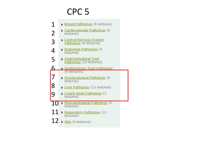
CPC 5 1 2 3 4 5 6 7 8 9 10 11 12
Case 1. A 27 year old woman attends her general practitioner for a routine cervical smear which is reported as severely abnormal (severe dyskaryosis). She attends colposcopy and a LLETZ is performed. Histology shows pleomorphic cells and numerous mitoses throughout the full thickness of the squamous epithelium. There is no breach of the basement membrane. What is the diagnosis? What is the cause of this condition? If untreated, what may develop? What term is given to precancer at the following anatomical sites? Large intestine Skin Breast Fallopian tube
Case 2. A 31 year old woman has dull left sided abdominal pain exacerbated by intercourse. A CT scan of abdomen and pelvis shows an enlarged solid and cystic ovarian mass. This is removed surgically and histology shows a variety of different mature tissue types such as cartilage, bone, teeth and skin. What is the diagnosis? Why is it important for the pathologist to take multiple samples from the tumour? A vary rare complication of this lesion is encephalitis. What is the underlying mechanism?
Case 3. A 39 year old woman has vague symptoms of abdominal bloating and increased urinary frequency. She complains of heavy periods. A hysterectomy is performed. Multiple fairly well circumscribed lesions are noted in the uterine wall. On sectioning the lesions have a white whorled appearance. Histology shows uniform spindle cells. There is no necrosis and no mitotic figures are seen. What is the probable diagnosis? Histology shows spindle cells. In general terms, what does the presence of spindle shaped cells normally signify? A 67 year old woman has a uterine tumour. Histology shows pleomorphic spindle cells, necrosis and several mitotic figures . What TWO differential diagnoses would be most likely?
Case 4. A 35 year old woman has an elective bilateral mastectomy and bilateral salpingoophorectomy. What is the most likely clinical indication for this operation? On examination of the ovaries, the pathologist looks carefully for any evidence of malignancy. What are the broad categories of ovarian malignancy? The ovaries are normal, but the pathologist notices that that there is severe nuclear abnormality of the epithelium of the fimbria / distal left fallopian tube with increased mitotic activity but no invasion. Discuss the significance of this abnormality.
Case 5. A 49 year old man is an inpatient in the regional liver unit. He has a history of cirrhosis due to Hepatitis C infection. On examination of the abdomen distended and engorged paraumbilical veins, which are seen radiating from the umbilicus. (Caput Medusa - Latin for "head of Medusa") - resembles Medusa's hair once Athena had turned it into snakes. Why is the abdomen distended. Give TWO mechanisms in your answer? Why are veins dilated on the anterior abdominal wall? At what other anatomical sites could distended veins also be present in this patient?
Case 6. A 36 year old woman has an ultrasound guided liver biopsy. The radiologist completes the pathology request form and provides the following clinical information. Deranged LFTs. Histopathology please. Many thanks++ Histology shows that within the liver tissue there are several rounded aggregates of macrophages with occasional multinucleated giant cells. What is the histological pattern? What is the clinical diagnosis? What information is missing from the clinical information?
Case 7. A 75 year old man presents to the GP feeling generally unwell and with episodes of abdominal pain . He is noted to be jaundiced. His liver feels enlarged. Blood tests reveal abnormal liver function tests. He had a blood transfusion 20 years previously after being involved in a road traffic accident in Uganda. He dies and at autopsy liver cirrhoisis is noted and in addition there is a primary liver tumour in keeping with hepatocellular carcinoma. What tumour marker is usually elevated in hepatocellular carcinoma? This tumour marker is also elevated in which other malignancy? What aetiological factors may cause this lesion?
Case 8. A 47 year old male presents to the GP feeling generally unwell. Blood tests reveal abnormal liver function tests and a raised serum ferritin. A liver biopsy is performed. Histology shows liver parenchyma with extensive deposition of brown pigment. A Perl s (iron) stain is positive. What is the diagnosis? What other organs may be affected? Why are men more severely affected than women? The patient tells you that he is planning to fly to Canada and asks if there are any special precautions he should take?
Cases 9 12 Lymph Node Pathology
Case 9. A 51 yr old woman has a 10 week history of painless enlarged lymph node in the left side of her neck. In broad terms, what are the potential causes of lymphadenopathy? The lymph node is excised and is reported as a follicular lymphoma. How are lymphomas categorised in general and in which category is follicular lymphoma? As well as routine H+E staining, which other stains are essential in the diagnosis of lymphoma?
Case 10. A 31 yr old woman presents with 4 week history of night sweats with weight loss. On examination enlarged, rubbery, mobile lymph nodes are noted in the axillae and neck. CT scan shows enlarged mediastinal lymph nodes. A lymph node is excised and histology shows scattered malignant cells with Owl s eye nuclei which are CD15+ and CD30+ in a background containing granulomas and lots of eosinophils What is the most likely diagnosis? The TNM staging system is a commonly used staging tool for carcinomas. Why is it not used in lymphoma? A different patient has pT3N1M0 colon cancer. One regional lymph node is involved by metastatic adenocarcinoma. How can the stage be M0 if there is a metastasis? https://openclipart.org/detail/17566/cartoon-owl
Case 11. You are working as an F2 doctor in ENT outpatients and a 27 year old man is being investigated for cervical lymphadenopathy. Two possible investigations for this patient are fine needle aspiration and excision of the lymph node. Explain the advantages and disadvantages of each procedure.
Ultrasound guided fine needle aspirate of lymph node: image c/o Dr E Napier
Cellular Pathology Cytopathology Histopathology
Case 12. A 46 year old man has a several month history of patches and plaques in a widespread distribution. A punch biopsy is reported as mycosis fungoides. Define mycosis fungoides. Several months later he presents with suspected Sezary syndrome. Explain what has happened to the patient. How does lymphoma differ from leukaemia?
