Hamartomas: A Case Study in Thoracic Medicine
Hamartomas, benign focal malformations resembling neoplasms, can arise in various body parts. This case study details the diagnosis, surgical intervention, and postoperative recovery of a large anterior mediastinal hamartoma in a previously healthy 33-year-old woman. Discover insights into the clinical presentation, histological features, and management of pulmonary hamartomas, one of the most common benign lung tumors.
Download Presentation

Please find below an Image/Link to download the presentation.
The content on the website is provided AS IS for your information and personal use only. It may not be sold, licensed, or shared on other websites without obtaining consent from the author.If you encounter any issues during the download, it is possible that the publisher has removed the file from their server.
You are allowed to download the files provided on this website for personal or commercial use, subject to the condition that they are used lawfully. All files are the property of their respective owners.
The content on the website is provided AS IS for your information and personal use only. It may not be sold, licensed, or shared on other websites without obtaining consent from the author.
E N D
Presentation Transcript
A hamartoma resembles a neoplasm in the tissue of its origin A developmental malformation Clonal chromosomal aberrations that are acquired through somatic mutations On this basis are now considered to be neoplastic Grows at the same rate as the surrounding tissue. It is composed of tissue elements normally found at that site, but they are growing in a disorganized manner. Hamartomas occur in many different parts of the body, most often asymptomatic incidentalomas (undetected until they are found incidentally on an imaging study obtained for another reason). hamartoma is a mostly benign, focal malformation that
A previously healthy 33-year-old lady was investigated for worsening breathlessness. Chest radiography demonstrated a mass in the left chest and a subsequent CT scan revealed a large anterior mediastinal mass extending mostly into the left hemithorax and compressing the left lung. CT guided bviopsy was s/o hamartoma. Surgical excision of this mass was indicated due to its large size.
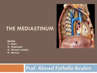
The patient underwent a left thoracotomy A giant fibrocartilagineous tumour was found arising superficially from the medial border of the left lung occupying most of the left chest and extending to the anterior mediastinum. The mass was compressing the left lung with no evidence of local invasion. The left phrenic nerve was identified and preserved, and the tumour was found to be adherent to the medial border of the lung and was easily dissected with sharp dissection and removed en bloc. left posterolateral posterolateral thoracotomy
The postoperative recovery was uneventful Patient was discharged home on the seventh postoperative day. Three months postoperatively the patient remains well. The final histology of the tumour, measuring 25.5 17.5 15.5 cm, and weighing 2200 g was a pulmonary hamartoma with predominantly adipose and cartilaginous differentiation with placental villi formation.
Most common form of benign lung tumours Incidence 0.025% 0.3% Parenchymal lesions more common, endobronchial lesions only about 10% The peak incidence is between the sixth and seventh decades with a male preponderance
The parenchymal lesions are usually an incidental radiological finding of a round homogeneous opacity in the periphery Endobronchial lesions are usually associated with haemoptysis and obstructive pneumonia . The size of these parenchymal lesions ranges from 1 8 cm and the tumour in our case is more than 3 times that of any previously published The histology of the parenchymal lesions usually reveals a predominant chondroid differentiation (80%), with fibroblastic (12%), fatty (5%) and osseous (3%) differentiation making the rest . In our case it was predominantly made of adipose and leiomyomatous tissue with placental villi differentiation. The size of the tumour and the histology make it an unusual presentation.
