Health and Technology in Brain Tumour Treatment: Advancements and Challenges
Brain tumours present challenges in detection and treatment due to their complex nature. Health professionals rely on imaging modalities such as MRI and CT scans for accurate diagnosis and segmentation. Advancements in artificial intelligence have enhanced the process of tumour detection, aiding in targeted therapies. However, challenges persist in accurate segmentation and classification of brain tumours for effective treatment planning.
Download Presentation

Please find below an Image/Link to download the presentation.
The content on the website is provided AS IS for your information and personal use only. It may not be sold, licensed, or shared on other websites without obtaining consent from the author.If you encounter any issues during the download, it is possible that the publisher has removed the file from their server.
You are allowed to download the files provided on this website for personal or commercial use, subject to the condition that they are used lawfully. All files are the property of their respective owners.
The content on the website is provided AS IS for your information and personal use only. It may not be sold, licensed, or shared on other websites without obtaining consent from the author.
E N D
Presentation Transcript
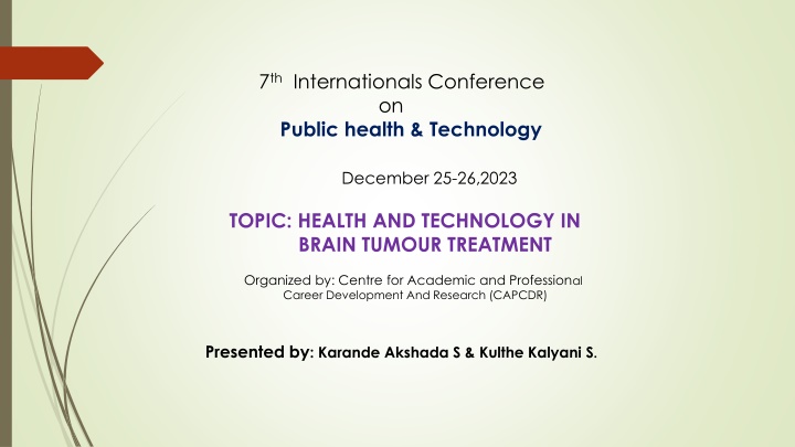
7th Internationals Conference on Public health & Technology December 25-26,2023 TOPIC: HEALTH AND TECHNOLOGY IN BRAIN TUMOUR TREATMENT Organized by: Centre for Academic and Professional Career Development And Research (CAPCDR) Presented by: Karande Akshada S & Kulthe Kalyani S.
HEALTH AND TECHNOLOGY IN BRAIN TUMOR TREATMENT A brain tumour is an uncontrolled and abnormal growth of brain cells. Any unexpected development can affect a person's functioning because the human skull is a rigid structure with a limited volume depending on the brain region involved. In addition, it can spread to other organs, further compromising human functions. Non-invasive techniques include physical inspections of the body and imaging modalities employed for imaging the brain In comparison to brain biopsy, other imaging modalities, such as CT scans and MRI images, are more rapid and secure. Radiologists use these imaging techniques to identify brain problems, evaluate the development of diseases, and plan surgeries . However ,brain scans or image interpretation to diagnose illnesses are prone to inter-reader variability and accuracy.
Brain tumour detection and segmentation is the most difficult and critical task for many medical imaging applications, as it often requires a large amount of information. Tumours come in different shapes and sizes. Automatic or semi-automatic detection/segmentation using artificial intelligence is now crucial in medical diagnosis. Before treatment, such as chemotherapy, radiation or brain surgery, medical professionals must identify the boundaries and areas of brain cancer and find out exactly where it is and what exact areas it affects. Brain tumours can be divided into several categories depending on the kind, place of origin, pace of development, and stage of progression; as a result, tumour classification is crucial for targeted therapy. Brain tumour segmentation aims to delineate accurately the areas of brain tumours.
Imaging modalities for brain tumour detection : MRI MRI is a non-invasive procedure that uses non-ionizing safe radiation to show the 3D anatomical structure of any body region without the need to cut tissue. It uses RF pulses and a strong magnetic field to produce images. The frame is designed to be placed in a strong magnetic field. The water molecules in the human body are initially in equilibrium when the magnets are turned off. The field is then activated by moving the magnets. The water molecules of the body align with the direction of the magnetic field under the influence of this strong magnetic field
2 CT-scan: (Computed Tomography ) CT scanners produce fine, detailed images of the inside of the body using a rotating x- ray beam and an array of detectors. Computer images taken from different angles are processed with special algorithms to create cross-sectional images of the whole body . However, a CT scan can provide more detailed images of the skull, spine, and other bony structures near the brain tumour, as shown in Figure 2. Patients usually receive contrast injections to highlight the abnormal tissue. A patient can sometimes take a dye to improve their image. If an MRI is not available and the patient has a pacemaker-like implant, a CT scan may be performed to diagnose a brain tumor. Advantages of CT scanning include low cost, better identification of tissue classification, rapid imaging, and wider availability. The radiation risk from a CT scan is 100 times greater than that of a standard X-ray diagnosis .
Brain tumor treatment using Radiation therapy: Radiation therapy can be given both inside and outside the body .The use of external beam radiation therapy A linear accelerator is a sizable device used in this treatment. High energy beams are directed by the machine to a specific point on the body. External beam radiation therapy uses a machine that directs high-energy beams into your body. This is called a linear accelerator. If you lie still, the linear acceleration will move around you. It emits radiation from several angles. Your care team has customized the machine just for you. In this way, it delivers a precise dose of radiation to a precise part of your body (25).You will not feel the radiation when it is delivered. It's like taking an x-ray. External beam radiation is an outpatient treatment. This means that you do not need to stay in the hospital after the treatment. Itcan common to get therapy five days a week over several weeks. Some treatment courses are given over 1 to 2 weeks. The treatment is spread out this way so that healthy cells have time to recover between sessions. Sometimes only one treatment is used to relieve pain or other symptoms from more advanced is the most popular.
Brain tumor treatment using Chemotherapy: Chemotherapy uses cancer (cytotoxic) drugs to kill brain tumour cells. Medicines circulate throughout the body in the bloodstream. You may receive chemotherapy after surgery or if the brain tumour comes back. Common chemotherapy drugs for brain tumours include a drug called temozolomide. And a combination of drugs called procarbazine, limestone and vincristine (PCV ). Brain tumours can be difficult to treat with some chemotherapy because the brain is protected by the blood-brain barrier. It is a natural filter between the blood and the brain that protects the brain from harmful substances. Figure 3. Chemotherapy uses drugs to kill cancer cells. This specific cancer treatment prevents cancer cells multiplying, dividing and producing new cells. Many cancers can be treated with chemotherapy. Your doctor may call chemotherapy standard chemotherapy. from
CONCLUSION: A brain tumour is an abnormal growth of brain tissue that affects the normal functioning of the brain. The main goal of medical image processing is to find accurate and useful information with as few errors as possible using algorithms. The four steps of brain tumour segmentation and classification using MRI data are pre-processing, image segmentation, feature extraction, and image classification. Automating the segmentation and classification of brain tumours can significantly improve diagnosis, treatment strategy and patient follow-up. Due to the appearance and irregular size, shape and nature of the tumour, it remains difficult to create a fully autonomous system that can be used in clinical layers. The main objective of the review is to present the most advanced imaging techniques in brain cancer, covering the pathophysiology of the disease.
REFERENCES: 1. DeAngelis L.M. Brain tumors. N. Engl. J. Med. 2001;344:114 123. Doi: 10.1056/NEJM200101113440207. [PubMed] [CrossRef] [Google Scholar 2. Hayward R.M., Patronas N., Baker E.H., V zina G., Albert P.S., Warren K.E. Inter-observer variability in the measurement of diffuse intrinsic pontine gliomas. J. Neuro-Oncol. 2008;90:57 61. doi: 10.1007/s11060-008-9631- 4. [PMC free article] [PubMed] [CrossRef] [Google Scholar] 3. Mahaley M.S., Jr., Mettlin C., Natarajan N., Laws E.R., Jr., Peace B.B. National survey of patterns of care for brain- tumor patients. J. Neurosurg. 1989;71:826 836. doi: 10.3171/jns.1989.71.6.0826. [PubMed] [CrossRef] [Google Scholar] 4. Sultan H.H., Salem N.M., Al-Atabany W. Multi-Classification of Brain Tumor Images Using Deep Neural Network. IEEE Access. 2019;7:69215 69225. doi: 10.1109/ACCESS.2019.2919122. [CrossRef] [Google Scholar. 5.Amyot F., Arciniegas D.B., Brazaitis M.P., Curley K.C., Diaz-Arrastia R., Gandjbakhche A., Herscovitch P., Hinds S.R., Manley G.T., Pacifico A., et al. A Review of the Effectiveness of Neuroimaging Modalities for the Detection of Traumatic Brain Injury. J. Neurotrauma. 2015;32:1693 1721. doi: 10.1089/neu.2013.3306. [PMC free article] [PubMed] [CrossRef] [Google Scholar] 6. Pope W.B. Brain metastases: Neuroimaging. Handb. Clin. Neurol. 2018;149:89 112. doi: 10.1016/b978-0-12- 811161-1.00007-4. [PMC free article] [PubMed] [CrossRef] [Google Scholar]
