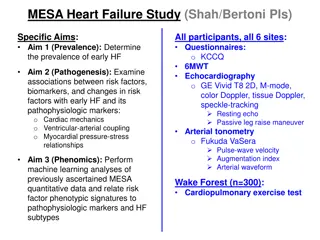
Heart Failure and Cardiovascular Dysfunction
Heart failure is a condition where the heart struggles to pump blood efficiently, leading to various symptoms and types of failure such as acute and chronic heart failure. Cardiovascular dysfunction can result from issues like filling defects, arrhythmias, or excessive workload, impacting the heart's ability to function effectively.

Download Presentation

Please find below an Image/Link to download the presentation.
The content on the website is provided AS IS for your information and personal use only. It may not be sold, licensed, or shared on other websites without obtaining consent from the author.If you encounter any issues during the download, it is possible that the publisher has removed the file from their server.
You are allowed to download the files provided on this website for personal or commercial use, subject to the condition that they are used lawfully. All files are the property of their respective owners.
The content on the website is provided AS IS for your information and personal use only. It may not be sold, licensed, or shared on other websites without obtaining consent from the author.
E N D
Presentation Transcript
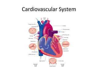
Diseases of the Cardiovascular Systeme
Functions of the cardiovascular system 1-Ensure an adequate circulation of blood so that nutrients are delivered, waste products are removed 2-a homeostatic environment will maintained at the organ and cellular level 3-Have a good role to maintain adequate circulation nutrient delivery and removal of waste product .
The two functional units of the cardiovascular system are the heart and the blood vessels; these two units are characterized as a pump (the heart) and a circuit (the blood vessels and blood).However, The pump and circuit may fail independently of each other, giving rise to two forms of circulatory failure, Heart failure and Circuit failure. Since, In heart failure the primary problem is inadequate pump performance, whereas in circuit failure the deficiency is in the vascular system.
Heart Failure And Cardio Vascular Dysfunction The failure of the heart as a pump can result from 1-A filling defect of the heart(diastolic failure) 2-Cardiac arrhythmia 3-An abnormality in the generation or conduction of the electrical wave of depolarization 4-An abnormality in contractile function(Contractile dysfunction) (systolic failure) 5-Excessive workload 6-Anatomic abnormalities 7- Obstructed flow 8- Regurgitant flow
Heart failure can divided into different types 1-Acute heart failure(AHF) . When cardiac output are in adequate result in an increase in venous pressure. there is an acute reduction of cardiac output, as is caused by sudden cessation of the heartbeat, the effect is to deprive tissues from oxygen supplies Moreover, itis a sudden, life-threatening condition in which the heart is unable to do its job. The heart is still beating, but it cannot deliver enough oxygen to meet the body's needs. Cardiac output . the quantity of blood pumped by the heart in a given period of time( Mostly one mint)
2-Chronic heart failure ( congestive heart failure)(CHF) . A condition in which the heart has trouble pumping blood through the body. It may develop over a long period of time. Symptoms include shortness of breath, problems exercising, fatigue, and swelling of the feet, ankles, and abdomen. Chronic heart failure may be caused by coronary artery disease, a heart attack, or high blood pressure. 3-Left side heart failure . It causes an increase in left ventricular end-diastolic pressure, which mean increase of left atrial pressure, and pulmonary venous pressure causing interstitial edema in the lungs and, severe enough, dyspnea, and death.
4- Right-sided heart failure . It causes an increase in right ventricular end-diastolic pressure, which mean increase in right atrial pressure, and jugular venous pressure causing symmetric venous distension (most readily detected in the jugular veins); an increase in pleural, pericardial, and abdominal fluid (ascites); and hepatomegaly.
Circuit Failure In circuit failure the effective blood volume is decreased because of 1-loss of fluid from the vascular system (hypovolemic shock) 2- by collection of blood in peripheral vessels 3-Increased capillary permeability (mald-distributive shock, which is commonly seen in endotoxemia). 4- The failure of venous return which results in incomplete filling of the heart and a reduction in cardiac output In general .. The effects of circuit failure are the same as those of chronic (congestive) heart failure because the supply of nutrients to the tissues and the removal of waste products from the tissues are reduced. .....
Cardiac Reserve And Compensatory Mechanisms In Heart Failure It could be done through 1-Increase in heart rate an elevation of heart rate alone is a significant factor in increasing cardiac output and complete filling of the heart 2-Increase in stroke volume Stroke volume is changeable and depends on the myocardial fibers Moreover, An increase in stroke volume is achieved primarily by an increase in the ejection fraction which depend on a-Filling pressure b-Contractile activity of myocardium c-The myocardial tension for ejection of blood d- The sequence of atrial and ventricular depolarization
3- Mixed Venous Oxygen Tension In normal animals at rest, the oxygen tension of mixed venous blood is above 40 mm Hg, When Stroke volume is reduced, the mixed venous oxygen tension falls below 40 mm Hg, reaching 15to 25 mm Hg resulting in severe shock and the arterial venous oxygen difference is large. There is also a redistribution of blood flow to vital organs 4- Autonomic Nerve Activity It is evident that increased sympathetic nerve activity also plays a significant role in compensating for the failing ventricle which act by increase heart rate and improving the contractility of the myocardium, and by increase venous return to the heart.
Measurement Of cardiac Reserve. It can be done by a- Assessment the heart rate b-Assessment of the intensity of the heart sounds c- The size of the heart d-The characteristics of the pulse e- The tolerance of the animal to exercise.
Cardiac Enlargement. We had to know that the weight of the heart is greater in athletic animals than in nonathletic animals. Moreover, the heart weight in horses can be modestly increased during training as a result of physiologic hypertrophy. In diseased animals The presences of cardiac enlargement on clinical examination is indicates the presence of a significant high flow volume load on the heart, due to the presence of myocardial disease and a reduction of cardiac reserve.
The enlargement of the heart is of two forms 1- Cardiac hypertrophy (concentric hypertrophy) Is the usual response to an increased pressure load, resulting in hypertrophy of individual heart fibers with an increase in the number of contractile units and an increase in total muscle mass and accompanied by decreased capillary density with increase inter-capillary distance . However, coronary blood flow will be limited. 2- Cardiac dilatation (eccentric hypertrophy) Is the usual response to an increased volume load results from fiber re-arrangement terminated with dilatation of cardiac champers which can eject a larger volume of blood as cardiac output
Special Examination of the Cardiovascular System 1-Examination of the heart .which include examination of Heart rate Heart rhythm Heart quality Abnormal heart sounds 2-Examination of arterial pulse and arterial blood pressure 3-Examination of Jugular vein 4-Percussion Of The Thorax To Identify The Cardiac Dullness Area 5-Exercise Tolerance
Manifestations of Circulatory Failure 1-Acute Heart Failure Etiology . 1-It occur due to severe defect in filling; when there is failure of the heart as a pump, caused by severe tachycardia, bradycardia and where there is a sudden increase in workload. 2-Severe excitement 3- Sudden onset of a severe arrhythmia, rupture of a heart valve or vessel, pericardial tamponade 4- Aortic and pulmonary artery rupture
5- Myocarditis, as in encephalomyocarditis virus, and FMD 6- Nutritional deficiency myopathy, as in copper or selenium deficiency 7- Plant poisoning, e.g., Phalaris spp., white snake root 8-Electrocution and lightning strike 9- Iatrogenic, as in IV calcium gluconate or borogluconate administration, or Xylazine. 10- Rupture of aortic valve 11-Acute anaphylaxis 12-Deep anesthesia
Clinical Findings. 1- The acute syndrome may occur while the animal is at rest but is most common during periods of excitement or activity 2- The animal usually shows dyspnea, staggering, and falling, and death often follows within seconds or minutes of the first appearance of signs. 3-Pallness of mucous membranes 4-Clonic convulsion with in coordination 5- weakness or absence of a palpable pulse and bradycardia, tachycardia, or absence of heart sounds 5- Death is usually preceded by deep, asphyxial gasps Treatment . Treatment of acute heart failure is not usually possible or practical in large animals because of the short course of the disease
2-Chronic (Congestive) Heart Failure Etiology . 1- Valvular Disease such as Valvular stenosis or valvular insufficiency Congenital valvular defects Rupture of valve 2- Myocardial Disease such as Myocarditis: bacterial, viral, parasitic, or toxic Myocardial degeneration: nutritional or toxic Congenital or hereditary cardiomyopathy Toxins affecting cardiac conduction
3- Congenital Anatomic Defects such as Cardiac defects, such as ventricular or atrial septal defects, tetralogy of Fallot Vascular abnormalities, patent ductus arteriosis 4- Hypertension such as Pulmonary hypertension: high altitude disease, cor pulmonale Systemic hypertension 5- Pressure Load It occur with lesions that produce an obstruction to outflow, such as aortic or pulmonary valve stenosis, during which the heart is required to perform more work to eject an equivalent amount of blood. 6-Volume Load Volume loads (flow loads) are common with both acquired and congenital heart defects. As In aortic valve and mitral valve insufficiency.
7-Pumping Defects (Systolic Failure) Myocarditis, cardiomyopathy, and neoplasms of the heart, are common causes of pumping defect 8-Filling Defects (Diastolic Failure) Pericardial diseases such as pericarditis and pericardial tamponade can result in diastolic filling Pathogenesis Congestive heart failure develops when the heart is unable to overcome on the circulatory requirement at rest.
Clinical Findings. 1-There is respiratory distress on light exercise with shortness of breath 2- problems exercising with fatigue 3- Swelling of the feet, ankles, and abdomen 4- pulse rates is prolonged with increase heart rate 5- There may be a loss of Body weight 6- subcutaneous edema (brisket edema commonly) and less frequently under the jaw (submandibular edema), However, Ascites could also seen in advance cases. all detect in right side congestive heart failure
7- The superficial veins are engorged and this is most easily detected by the presence of bilateral jugular vein distension 8- Hydrothorax and hydropericardium may also be clinically detected by auscultation 9-Enlargemnt of liver due to engorgement with blood 10- Urine flow is usually reduced and the urine is concentrated and contains a small amount of protein. 11- A cardiac cough associated with pulmonary edema with crackles sound detected in left side congestive heart failure
Treatment .. 1-Diuretics such as furosemide(0.25 to 1 mg/kg for horses and 2.5 to 5 mg/kg for cattle I.V) acetazolamide, or chlorothiazide is an important component of treatment because eliminates excess body fluids. 2- Angiotensin-Converting Enzyme Inhibitors such as Single oral doses of benazepril (0.5 mg/kg) was a more effective ACE inhibitor in healthy adult horses than single oral doses of ramipril (0.3 mg/kg),in cattle 3-The use of inotropic agent Such as Dobutamine IV doses (2.5 7.5 [ g/ kg]I M Digoxin IV dose of 0.01 to 0.015 mg/kg BW Im Calcium gluconate, calcium borogluconate, and calcium chloride S.C
3-Arrhythmias (Dysrhythmias) Its irregularity in rate and rhythm which influenced by the integrity of the pacemaker, the conducting system, the myocardium, and also by the influence of the autonomic nervous system Moreover, acid base and electrolytes disturbance can also affects, However, the treatment were always depend on the prevention of the primary cause . 4-Sinus Tachycardia(tachycardia) It s an increase in heart rate caused by detectable influences such as pain, excitement, exercise, hyperthermia, a fall in arterial blood pressure, or the administration of adrenergic drugs. The heart rate returns to normal when the influence is removed or relieved.
5-Sinus Bradycardia(Bradycardia) It s a decrease in heart rate most commonly associated with highly trained, and association with an increase in arterial blood pressure, space-occupying lesions of the cranium and increased intracranial pressure,pituitary abscess, hyperkalemia hypothermia, and hypoglycemia and following the administration of xylazine or detomidine. Bradycardia is sometimes associated with vagus indigestion and diaphragmatic hernia. 6- Atrial Fibrillation Is characterized by numerous independent appearance of excitation that continous and randomly through the atria accompanied by irregular irregularly of the heart and pulse rate result from atrial enlargement, However, in cattle it could also occur due to gastrointestinal diseases and metabolic diseases and treated mostly with Digoxin and quinidine sulfate
Diseases of the Heart Valvular Disease And Murmurs Etiology . 1- Endocarditis 2-Laceration, detachment and dilatation of aortic valve 3-Congenital Pulmonic valve stenosis. 4-Congenital Blood cysts are common on the aortic valve 5- mitral and tricuspid valve endocardiosis Pathogenesis Stenosis of the outflow valves results in an increased pressure load on the heart and compensatory hypertrophy. The important clinical indications of valvular disease are audible murmurs and palpable precordial thrills. Murmurs may occur at any phase of the cardiac cycle and are caused by the vibrations of fast flow of blood transmitted to the surface of the chest.
Clinical Findings 1- The clinical findings in chronic (congestive) heart failure will present here 2-Ascultation of cardiac murmurs either systolic, diastolic , continous ,intermittent and harsh Treatment . There is no specific treatment for valvular disease, although replacement of the disused valve some time advised in human .
Endocarditis Etiology . Bacterial infection ( in all animals )causes by Corynebacterium pyogenes , Actinobacillus equuli Helcococcus ovis, -Hemolytic streptococci, Pasteurella Micrococcus and Staphylococcus spp., E. coli Pseudomonas spp.,Clostridium chauvoei (blackleg) Mycoplasma mycoides
Pathogenesis Endocarditis may arise from implantation of bacteria onto the endocardium from the bloodstream or by bacterial embolism of the valve capillaries, It will predisposed by trauma to the endothelial surface exposing collagen and leading to binding of platelets, activation of the coagulation factors with deposition of fibrin, and the formation of sterile platelet fibrin deposits result in severe damage of the endocardial tissue causing infarction and affect the heart functions terminated with cardiac insufficiency and heart failure will terminated .
Clinical findings . 1-The important finding is a murmur on auscultation or a thrill on palpation of the cardiac area. 2- Persistent tachycardia is a clear clinical signs in animals 3- fluctuating fever, and secondary involvement of other organs may cause the appearance of signs of peripheral lymphadenitis, embolic pneumonia, nephritis, arthritis, tenosynovitis, or myocarditis. There is usually much loss of condition, pallor of mucosa. 4-Acute heart failure will finally cause death Treatment Treatment is not highly successful because of the difficulty in controlling the infection.
Traumatic pericarditis Its inflammation of pericardial membrane or sac of the heart and occur in 3 general forms Effusive ,Fibrinous and Constrictive Effusive is characterized by the accumulation of a protein rich fluid within the pericardial sac. Subsequent fibrin deposition can lead to fibrinous pericarditis, and if fibrin within the pericardial sac develop to fibrous tissue then fibrosis will occur of the pericardium or epicardium then constrictive pericarditis will result.
Etiology 1- Traumatic pericarditis, due to perforation of the pericardial sac by an infected foreign body,occurs commonly only in cattle. 2- Direct extension of infection from pleurisy or myocarditis may also occur in all animals. 3-Infection by Mannheimia hemolytica, Haemophilus spp, Pseudomonas aeruginosa, Mycoplasma spp. Klebsiella pneumoniae and Actinobacillus spp. Pathogenesis 1-In the early stages, inflammation of the pericardium is accompanied by hyperemia and the deposition of fibrinous exudates, which produces a friction sound when the pericardium and pericardium rub together during cardiac movement.
2-As effusion develops the inflamed surfaces are separated, thus the friction sound is replaced by muffling of the heart sounds 3- When accumulation of fluid occur it will compresses the atria and right ventricle, preventing their complete filling, therefore Congestive heart failure follows. 4- A severe toxemia is usually present in suppurative pericarditis because of the toxins produced by the bacteria in the pericardial sac. Gas will occur along with fluid in the sac if gas-producing bacteria are present. 5- In the chronic stage of non suppurative pericarditis the fluid is reabsorbed and adhesion will develop between the pericardium and epicardium to cause an adhesive pericarditis, but the adhesions are usually not sufficiently strong to impair cardiac movement
Clinical findings 1-In the early stages there is pain, characterized by avoidance of movement, abduction of the elbows, arching of the back and shallow abdominal respiration. Pain is evidenced on percussion or firm palpation over the cardiac area of the chest wall, 2-The animal lies down carefully. 3-A pericardial friction sound is detectable on auscultation of the cardiac area.
4-The body temperature is elevated to 39.5-41C and the pulse rate is increased. Associated signs of pleuritis, pneumonia and peritonitis may be present. 5-The second stage of effusion is manifested by muffling of the heart sounds and increase in the area of cardiac dullness with decreased amplitude of the peripheral pulse. If gas is present in the pericardial sac each cardiac cycle may be accompanied by splashing sounds then 6-death usually occur due to congestive heart failure or toxemia .
Diagnosis Can be done by 1-History and clinical sings 2- Clinical pathological examination revealed A marked leukocytosis and neutrophilia, as well as hyperglobulinemia, 3-Examination of Pericardial fluid 4- Electrocardiography 5- Radiography 6- Echocardiography
Myocardial diseases Its inflammation and / or degenerative disorders of the myocardial tissue. A number of diseases are accompanied by inflammation, necrosis, or degeneration of the myocardium. These include several bacterial, viral, or parasitic infections and some nutritional deficiencies and toxicities. Etiology .. 1-Following bacteremia, as in strangles or from navel-ill 2-Tuberculosis,and in Tick pyemia in lambs 3-Clostridium chauvoei, Histophilus somni in feedlot cattle 4-Extension from pericarditis, epicarditis, or endocarditis 5-FMD, African horse sickness, Equine infectious anemia, Equine herpesvirus-1 in fetus, Equine viral arteritis
6- Parasitic Myocarditis, This is primarily associated with Strongylus spp. (migrating larvae) cysticercosis, Sarcocystis spp., and Neospora caninum 6- Nutritional Deficiency, as in Vitamin E/selenium, and Some forms of chronic copper deficiency in cattle (falling disease). 7- Toxicity, such as Inorganic poisons: selenium, arsenic,mercury, phosphorus, Mycotoxicosis, some toxic plants and weeds,toxic drugs such as Xylazine and catecholamine, Snack bite venom. 8-Tumors and inherited diseases .
Pathogenesis.. 1-The primary effect of any myocardial lesion is to reduce cardiac reserve and limit compensation in circulatory emergencies. 2- myocardial disease results in arrhythmias and conduction disturbances 3- Myocardial disease may also result in congestive heart failure through its primary effect on the myocardium and the function of the heart as a pump.
Clinical Findings 1- Decreased exercise tolerance 2-Increase in heart rate and heart size, which can detectable by echocardiography. 3- There may be clinically recognizable arrhythmia 4- The characteristics of the pulse and heart sounds are also changed 5-Sudden death or attacks of cardiac syncope caused by acute heart failure, or severe dyspnea or general edema caused by congestive heart failure. Treatment Treatment of the primary cause is advised
Cor Pulmonale Is a condition that causes the right side of the heart to fail. Long-term high blood pressure in the arteries of the lung and right ventricle of the heart can lead to cor pulmonale. Etiology 1- alveolar hypoxia 2-Animal living at high altitude areas (high mountain disease) 3- Chronic interstitial pneumonia and emphysema
High Altitude Pulmonary Hypertension (Brisket Disease), (Mountain Sickness).. Etiology .. 1-Alveolar hypoxia in cattle at high altitudes results in pulmonary hypertension 2- Myocardial dystrophy, anemia, pulmonary disease, or hypoproteinemia Pathogenesis . 1- Acute alveolar hypoxia (lowered alveolar Po2) 2- Prolonged hypoxia and persistent pulmonary vasoconstriction lead to medial muscular hypertrophy of the small pulmonary arteries and arterioles with a further increase in pulmonary vascular resistance and the development of cor pulmonale
Clinical Findings.. 1- Affected cattle have a dejected appearance; lose condition rapidly; have a rough, lusterless coat; and stand with the elbows abducted. 2- Jugular vein engorgement is followed by the appearance of edema of the brisket, spreading up the neck to the inter-mandibular space and back along the ventral aspect of the body. 3- Abdominal enlargement caused by the development of ascites is accompanied by diarrhea. 4- There is dyspnea and weakness on slight exertion. The mucosae may be cyanotic, particularly after exercise, and the lung sounds vary from an increased vesicular murmur to moist crackles and an absence of breath sounds. 5- Auscultation of the heart reveals tachycardia, increased absolute intensity of the sounds Treatment The only effective treatment is to move the affected cattle to a lower altitude


 Heart Failure")

")


")
")