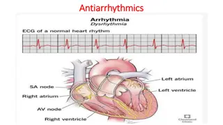
Heart Rate Variability and Arrhythmias in Thoroughbred Racehorses
Beat-to-beat heart rate variations, HRV as a prognostic indicator for cardiac health, detection of arrhythmias during exercise, exploring associations with clinical conditions in Thoroughbred racehorses.

Download Presentation

Please find below an Image/Link to download the presentation.
The content on the website is provided AS IS for your information and personal use only. It may not be sold, licensed, or shared on other websites without obtaining consent from the author.If you encounter any issues during the download, it is possible that the publisher has removed the file from their server.
You are allowed to download the files provided on this website for personal or commercial use, subject to the condition that they are used lawfully. All files are the property of their respective owners.
The content on the website is provided AS IS for your information and personal use only. It may not be sold, licensed, or shared on other websites without obtaining consent from the author.
E N D
Presentation Transcript
Heart rate variability and arrhythmias in Thoroughbred racehorses undergoing treadmill exercise and recovery. WILLIAM SAGE CONTRIBUTORS: KATE ALLEN, MELANIE HEZZELL AND ANNA HAMMOND
HEART RATE VARIABILITY (HRV) Beat-to-beat heart rate variations Neurohormonal control HR is dynamic Time-domain measures offer overall assessment of autonomic function SDRR: simplest indicator of R-R variability RMSSD: estimate of high-frequency fluctuations Frequency-domain measures VLF/ULF (very-low/ultra-low): renin-angiotensin-aldosterone system and posture (seconds to minutes) LF (low): sympathetic nervous system (seconds) HF (high): parasympathetic nervous system on a beat-to-beat basis Entropy values Measures of irregularity in a biological system
Why? HRV has been used as a prognostic indicator for cardiac health at rest in humans and horses There is a growing interest in using HRV for the detection of arrhythmias during exercise Arrhythmias are commonly observed during exercising Thoroughbreds during treadmill exercise Can we validate the use of HRV analysis during exercise and find useful associations with clinical conditions, including arrhythmias?
Literature Physick-Sheard P.W., et. al., (2000) Frequency domain analysis of heart rate variability in horses at rest and during exercise Physick-Sheard P.W. and McGurrin M.K. (2010) Ventricular arrhythmias during race recovery in standardbred racehorses and associations with autonomic activity Frick L., et. al., (2019) The use of heart rate variability analysis to detect arrhythmias in horses undergoing a standard treadmill exercise test Massie S.L., et. al., (2021) Prevalence of cardiac arrhythmias and R-R interval variation in healthy Thoroughbred horses during official Chuckwagon races and recovery
Objectives Report HRV indices during submaximal exercise, strenuous exercise and recovery and explore associations with arrhythmia in Thoroughbred (TB) racehorses. Investigate relationships between HRV indices and other common causes of poor performance in TB racehorses including; lameness, gastric ulceration (EGUS), lower airway inflammation and upper respiratory tract (URTO) obstructions.
Study Design Clinical records of TB racehorses, which had presented for high- speed treadmill investigation of poor performance between 2007 and 2018 were retrospectively reviewed to facilitate a prospective analysis of associations between HRV, arrhythmias and clinical conditions. All horses underwent a full clinical examination, including an assignment of a lameness grade, upper airway endoscopy and electrocardiography (ECG) during the treadmill test. Some horses underwent further clinical investigations, including; lower airway evaluations (tracheal endoscopy, tracheal wash (TW), bronchoalveolar lavage (BAL)) and/or gastroscopy.
Treadmill exercise test (SIET) Sub-maximal phase 5% incline for 2 minutes at walk (1.8m/s), 4 minutes at trot (3.5m/s) 1 minute at canter (6 or 7 m/s). Strenuous phase 1 min at each of 6, 8 and 10 m/s on a 10% incline Further increments of 1 m/s at 1 min intervals. Recovery phase Horses were walked on the treadmill on a 0% incline for 5 minutes From 2011, a high-speed test (HSET) was introduced for horses in training for flat racing consisting of 1 min at each of 11 and 12m/s on a 10% incline.
Methods Self-adhesive electrodes were placed in a modified base-apex configuration Recordings were obtained and analysed using a telemetric ECG system (Televet 100) Recordings were corrected manually to ensure correct R wave detection No ectopic beats or arrhythmias were deleted from the recordings Text files of the R-R intervals were imported into HRV software (Kubios 3.2) Results were calculated from the non- detrended R-R series and all Kubios filters were removed
Methods Strenuous exercise Recovery phase Submaximal exercise
Arrhythmia Arrhythmias were recorded separately during submaximal exercise, strenuous exercise and recovery Horses were categorised into two groups, either with or without arrhythmia Horses were assigned to the arrhythmia groups if they had any occurrence of non-sinus rhythm, including: supraventricular or ventricular premature depolarisations sinus arrhythmia atrioventricular block paroxysmal atrial fibrillation. An 8% cut-off was applied to determine truly premature events, along with visual operator assessment
Arrhythmia SVPD couplet followed by isolated VPD in recovery
HRV analysis Kubios:
Statistical analysis The data were analysed using IBM SPSS Distribution and variance of the HRV measures were tested for normality using Kolmogorov-Smirnov tests and visual inspection of histograms Univariate linear regressions were constructed to assess associations between each HRV measure and the clinical variables Variables associated at the 20% level were then entered into a backwards, stepwise multivariable linear regression model The residuals were inspected visually to assess for normality and the variables were transformed appropriately when required Significance was set at P<.05
Results Frequency Frequency Variable Variable (Yes / No / missing data) (Yes / No / missing data) Lameness 62 / 47 / 71 Upper airway obstruction (URTO) 128 / 51/ 1 Lower airway inflammation 74 / 40 / 66 Gastric ulceration (EGUS) 58 / 26 / 96 High-speed exercise test (HSET) 12 / 168 / 0 Arrhythmia during submaximal exercise Arrhythmia during submaximal exercise 10 / 166 / 4 10 / 166 / 4 Arrhythmia during strenuous exercise Arrhythmia during strenuous exercise 71 / 109 / 0 71 / 109 / 0 Arrhythmia during recovery Arrhythmia during recovery 131 / 49 / 0 131 / 49 / 0
Time-domain Results SDRR in strenuous exercise RMSSD in strenuous exercise Variable Variable B B 95% CI for B 95% CI for B P P Variable Variable B B 95% CI for B 95% CI for B P P Lameness -.046 -.129 to .037 .271 Lameness -.043 -.119 to .033 .263 URTO -.004 -.074 to .066 .906 URTO -.004 -.069 to .061 .907 Lower airway inflammation -.022 -.099 to .054 .562 Lower airway inflammation -.009 -.080 to .063 .811 EGUS .035 -.058 to .129 .453 EGUS .033 -.055 to .121 .456 HSET .071 -.056 to .198 .273 HSET .074 -.044 to .192 .215 Time in strenuous exercise .000 .000 to .001 .353 Time in strenuous exercise .000 .000 to .001 .381 Arrhythmia during strenuous exercise Arrhythmia during strenuous exercise .238 .183 to .293 <.001 .221 .171 to .272 <.001
Time-domain Results SDRR in recovery RMSSD in recovery (A) (A) Variable Variable B B 95% CI for B 95% CI for B P P (A) (A) Variable Variable B B 95% CI for B 95% CI for B P P Lameness .063 -.102 to .229 .455 Lameness .058 -.093 to .209 .447 URTO .080 -.065 to .224 .278 URTO .090 -.042 to .222 .182 Lower airway inflammation -.047 -.207 to .112 .559 Lower airway inflammation -.042 -.189 to .105 .572 EGUS .025 -.172 to .223 .799 EGUS .009 -.177 to .194 .925 HSET .048 -.213 to .309 .717 HSET .088 -.151 to .327 .469 Time in strenuous exercise .002 .000 to .003 .013 Time in strenuous exercise .001 .000 to .003 .042 Arrhythmia during recovery .614 .500 to .729 <.001 Arrhythmia during recovery .498 .386 to .610 <.001 (B) Variable B 95% CI for B P (B) Variable B 95% CI for B P TE .001 .000 to .002 .031 Arrhythmia during recovery .512 .400 to .624 <.001 Arrhythmia during recovery .604 .490 to .718 <.001
Frequency-domain Results VLF in strenuous exercise VLF in strenuous exercise HF in recovery phase HF in recovery phase (A) (A) Variable Variable B B CI CI P P (A) (A) Variable Variable B B CI CI P P Lameness 1.415 -6.004 to 8.834 .706 Lameness -.204 -.456 to .048 .112 URTO 1.874 -4.569 to 8.317 .567 URTO -.176 -.380 to .028 .090 Lower airway inflammation 2.175 -5.402 to 9.751 .571 Lower airway inflammation -.000 -.234 to .234 >.999 EGUS 2.917 -6.315 to 12.149 .531 EGUS -.090 -.373 to .194 .530 HSET -7.087 -18.661 to 4.487 .228 HSET -.171 -.541 to .199 .363 Time in strenuous exercise .093 .027 to .159 .006 Time in strenuous exercise -.001 -.003 to .002 .632 Arrhythmia during strenuous exercise Arrhythmia during recovery 11.041 4.737 to 17.346 <.001 -.278 -.463 to .093 .003 (B) Variable B CI P (B) Variable B CI P Time in strenuous exercise .084 .019 to .148 .012 URTO -.274 -.553 to .006 .055 Arrhythmia during strenuous exercise Arrhythmia during recovery 10.337 4.103 to 16.570 .001 -.292 -.548 to -.036 .026
Discussion Time domain HRV HRV measures in exercise were lower and within a tighter range than HRV measures in recovery Arrhythmogenesis was highest in recovery period All time-domain HRV parameters were increased in horses with arrhythmias (barring log SDRR in submaximal period) Lame horses had a decreased log RMSSD during submaximal exercise During recovery, arrhythmia increased all time-domain measures
Discussion Frequency domain HRV During submaximal exercise, lower airway inflammation and EGUS influenced frequency domain HRV measures During strenuous exercise, URTO and EGUS influenced frequency domain HRV measures During recovery, arrhythmia and time in exercise influenced all frequency domain HRV measures
Limitations Arrhythmia identification and classification Cut-off points of clinical conditions for statistical analysis Spectrum of severity
Conclusions Arrhythmia influenced HRV measures in all phases of exercise and recovery And Lameness, lower airway inflammation and HSET influenced HRV measures in submaximal exercise EGUS and URTO influenced HRV measures in strenuous exercise Time in exercise influenced HRV parameters in recovery phase
References 1. Allen, K.J., Young, L.E. and Franklin, S.H. (2016) Evaluation of heart rate and rhythm during exercise , Equine Veterinary Education, 28, pp. 99-112. 2. Broux, B., De Clercq, D., Decloedt, A., Ven, S., Vera, L., van Steenkiste, G., Mitchell, K., Schwarzwald, C., and van Loon, G. (2017) Heart rate variability parameters in horses distinguish atrial fibrillation from sinus rhythm before and after successful electrical cardioversion , Equine veterinary journal, 49(6), pp. 723 728. 3. da Silva, V. P., de Oliveira, N. A., Silveira, H., Mello, R. G., and Deslandes, A. C. (2015) Heart rate variability indexes as a marker of chronic adaptation in athletes: a systematic review , Annals of Noninvasive Electrocardiology , The Official Journal of the International Society for Holter and Noninvasive Electrocardiology, 20 (2), pp. 108 118. 4. Frick, L., Schwarzwald, C.C. and Mitchell, K.J. (2019) The use of heart rate variability analysis to detect arrhythmias in horses undergoing a standard treadmill exercise test , Journal of Veterinary Internal Medicine 33, pp. 212-224. 5. G sior, J. S., Hoffmann, B., Silva, L., Ma ek, ., Flatt, A. A., Baranowski, R. and Werner, B. (2020) Changes in Short-Term and Ultra-Short Term Heart Rate, Respiratory Rate, and Time-Domain Heart Rate Variability Parameters during Sympathetic Nervous System Activity Stimulation in Elite Modern Pentathlonists - A Pilot Study , Diagnostics, 10 (12), p. 1104. 6. Marr, C.M., Franklin, S., Garrod, G., Wylie, C., Smith, L., Dukes-McEwan, J., Bright, J. and Allen, K. (2020) Exercise-associated rhythm disturbances in poorly performing Thoroughbreds: risk factors and association with racing performance , Equine Veterinary Journal, 53, pp. 656-669. 7. Massie, S.L., Bezugley, R.J., McDonald, K.J., Leguillette, R. (2021) Prevalence of cardiac arrhythmias and R-R interval variation in healthy Thoroughbred horses during official Chuckwagon races and recovery , The Veterinary Journal, 267, pp. 1090-0233.
References continued 8. Michael S., Graham K.S., Davis G.M. OAM. (2017) Cardiac Autonomic Responses during Exercise and Post-exercise Recovery Using Heart Rate Variability and Systolic Time Intervals A Review . Frontiers in Physiology. 29, pp. 8-301. 9. Physick-Sheard P.W. and McGurrin M.,K. (2010) Ventricular arrhythmias during race recovery in standardbred racehorses and associations with autonomic activity . Journal of Veterinary Internal Medicine, 24, pp. 1158 66. 10. Physick-Sheard P.W., Marlin D.J., Thornhill R. and Schroter R.C. (2000) Frequency domain analysis of heart rate variability in horses at rest and during exercise , Equine Veterinary Journal, 32(3), pp. 253-62. 11. Ryan N., Marr C.M. and McGladdery A.J. (2005) Survey of cardiac arrhythmias during submaximal and maximal exercise in thoroughbred racehorses , Equine Veterinary Journal, 37, pp. 265 8. 12. Schmidt A., Biau S., M stl E., Becker-Birck M., Morillon B., Aurich J., Faure J.M. and Aurich C. (2010) Changes in cortisol release and heart rate variability in sport horses during long-distance road transport , Domestic Animal Endocrinology, 38(3), pp. 179-89. 13. Shaffer F. and Ginsberg J.P. (2017) An Overview of Heart Rate Variability Metrics and Norms , Frontiers in Public Health, 5, p. 258. 14. Slack, J., Stefanovski, D., Madsen, T.F., Fjordbakk, C.T., Strand, E. and Fintl, C. (2021) Cardiac arrhythmias in poorly performing Standardbred and Norwegian Swedish Coldblooded trotters undergoing high-speed treadmill testing , The Veterinary Journal, 267. 15. Vitale, V., Viu, J., Armengou, L., R os, J. and Jose-Cunilleras, E. (2020) Prognostic value of measuring heart rate variability at the time of hospital admission in horses with colic , American journal of veterinary research, 81(2), pp. 147 152.
Thank you for listening ANY QUESTIONS?

")


")