Heart Sounds: Normal S1 and S2
Heart sounds, including S1 (LUB) and S2 (DUB), are generated by valve closures in the heart. Abnormal sounds like S3 and S4 may indicate heart problems like myocardial failure or ventricle overfilling. Learn to differentiate between normal and abnormal heart sounds using auscultation techniques.

Download Presentation

Please find below an Image/Link to download the presentation.
The content on the website is provided AS IS for your information and personal use only. It may not be sold, licensed, or shared on other websites without obtaining consent from the author.If you encounter any issues during the download, it is possible that the publisher has removed the file from their server.
You are allowed to download the files provided on this website for personal or commercial use, subject to the condition that they are used lawfully. All files are the property of their respective owners.
The content on the website is provided AS IS for your information and personal use only. It may not be sold, licensed, or shared on other websites without obtaining consent from the author.
E N D
Presentation Transcript
AUSCULTATION AUSCULTATION HEART SOUND
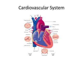
NORMAL HEART SOUND (S1 & S2) 1- The first heart sound LUB is produced by the closing of the mitral and tricuspid valve leaflets. 2-The second heart sound DUB is produced by the closing of the aortic and pulmonic valve leaflets.
NORMAL HEART SOUND (S1 & S2) Pulmonary valve Aortic valve Apex of the heart Tricuspid valve Click below for audio Click below for patient recording
S3 SOUND Cause of S3 : It Is produced when blood from atria (atrium) falls into ventricles in a diastole. The rapid flow of blood causes vibration of the resulting from the first rapid filling so it is heard just after S2. ventricular walls,
S3 SOUND Flow of blood from Atrium into Ventricle
S3 SOUND S3 is considered normal in young children. But in adults more than 40 yrs in age S3 sound may indicate heart problems. An abnormal S3 is associated with: Decreased intrinsic ability of the heart to contract known as decreased myocardial contraction Heart failure also known as myocardial failure Overfilling of ventricles with blood also known as volume overload of a ventricle because of mitral or tricuspid regurgitation (backflow of blood)
S3 SOUND Chespiece : Diaphragm of stethoscope Location of stethoscope: On the left : Apex of the heart On the right : At the tricuspid region.
S3 SOUND Apex of the heart Tricuspid valve Click below for audio Click below for patient recording
S4 SOUND S4 sound is produced when blood flows forcefully into a stiffened ventricle/ventricles due to active contraction of the atria. It is heard when the diastole is about to end and systole is about to begin. So it s heard just before S1.
S4 SOUND Flow of blood from Atrium into Ventricle due to contraction
S4 SOUND An S4 sound may indicate An S4 sound may indicate: : Myocardial Myocardial "myocardial the myocardium (the heart muscle) and the changes that occur in it due to the sudden deprivation of circulating blood. The main change is necrosis (death) of myocardial tissue.) The word "infarction" comes from the Latin "infarcire" meaning "to plug up or cram." It refers to the clogging of the artery. ischemia/infarction ischemia/infarction infarction" (The term on focuses
S4 SOUND LV LV hypertrophy hypertrophy Left ventricular hypertrophy is enlargement (hypertrophy) of the muscle tissue that makes up the wall of your heart's main pumping chamber (left ventricle). Left ventricular hypertrophy develops in response to some factor, such as high blood pressure, that requires the left ventricle to work harder. As the workload increases, the walls of the chamber grow thicker, lose elasticity and eventually may fail to pump with as much force as that of a healthy heart.
S4 SOUND Chespiece : Diaphragm /bell of stethoscope Location of stethoscope: On the left : Apex of the heart On the right : At the tricuspid region.
S4 SOUND Apex of the heart Tricuspid valve Click below for audio Click below for patient recording
SYSTOLIC EJECTION MURMUR Sound produced when the blood is unable to flow freely from ventricle into the aorta due to stiffness in the aortic valve.
SYSTOLIC EJECTION MURMUR Stiff & Narrow Aortic Valve
SYSTOLIC EJECTION MURMUR Systolic ejection murmur may indicate : Systolic ejection murmur may indicate : Aortic Aortic Stenosis Stenosis : : In aortic stenosis, the aortic valve does not open fully due to calcium deposits that narrow the valve.
SYSTOLIC EJECTION MURMUR Chestpiece : Diaphragm Location of stethoscope: Right Upper Sternal Border.
SYSTOLIC EJECTION MURMUR Aortic Valve Click below for audio Click below for patient recording
HOLOSYSTOLIC MURMUR HOLOSYSTOLIC MURMUR Sound produced due to backflow of blood from ventricles back into the atrium due to malfunctioning of mitral valve.
HOLOSYSTOLIC MURMUR HOLOSYSTOLIC MURMUR Back flow of blood from Ventricles into Atrium due to Mitral valve malfunction
HOLOSYSTOLIC MURMUR HOLOSYSTOLIC MURMUR Holosystolic Holosystolic murmur may indicate: murmur may indicate: Mitral Regurgitation due to mitral valve prolapse. i.e backflow of blood into the atrium when mitral valve malfunctions.
HOLOSYSTOLIC MURMUR HOLOSYSTOLIC MURMUR Chespiece Chespiece : : Diaphragm Location of stethoscope: Location of stethoscope: Mitral valve area
HOLOSYSTOLIC MURMUR HOLOSYSTOLIC MURMUR Mitral Valve Click below for audio Click below for patient recording
EARLY DIASTOLIC MURMUR EARLY DIASTOLIC MURMUR Sound produced due to backflow of blood from aorta (major artery carrying blood to the body) and pulmonary artery( artery carrying blood to the lungs) back into the ventricles.
EARLY DIASTOLIC MURMUR EARLY DIASTOLIC MURMUR Back flow of blood from Aorta into Ventricle
EARLY DIASTOLIC MURMUR EARLY DIASTOLIC MURMUR Early Early diastolic diastolic murmur Aoritc Aoritc regurgitation regurgitation : Backflow of blood from from aorta into ventricles when the valve doesnot close properly. . Pulmonary Pulmonary regurgitation regurgitation: : backflow of blood from pulmonary artery back into ventricle when the pulmonary valve doesnot close properly. murmur may may indicate indicate: :
EARLY DIASTOLIC MURMUR EARLY DIASTOLIC MURMUR Chespiece Chespiece : : Diaphragm Location of Location of stethoscope: stethoscope: Aortic and Pulmonary valve
EARLY DIASTOLIC MURMUR EARLY DIASTOLIC MURMUR Pulmonary Valve z Aortic Valve Click below for audio Click below for patient recording
MID SYSTOLIC CLICK (Heart murmur) It is heard when Mitral Valve doesn t close properly.
MID SYSTOLIC CLICK (Heart murmur) Half closed mitral valve
MID SYSTOLIC CLICK (Heart murmur) Midsystolic click may indicate MITRAL VALVE PROLAPSE which means Mitral valve is not able to close properly. This is because the connecting tissue of the valve which allows it to open and close properly has degenerated.
MID SYSTOLIC CLICK (Heart murmur) Chespiece : Diaphragm Location of stethoscope: Mitral valve area
MID SYSTOLIC CLICK (Heart murmur) Mitral Valve Click below for audio Click below for patient recording
INNOCENT SYSTOLIC MURMUR Is produced due to flow of from ventricles into the aorta (major artery supplying blood to the body) or the pulmonary artery( artery taking blood to the lungs) .
INNOCENT SYSTOLIC MURMUR From Ventricle into Aorta From Ventricle into Pulmonary Artery
INNOCENT SYSTOLIC MURMUR Innocent Innocent murmer Defective heart valves Pregnancy, Fever, Hyperthyroidism (Overactive Thyroid Gland) Anemia. murmer may indicate may indicate
INNOCENT SYSTOLIC MURMUR Chespiece : Diaphragm Location of stethoscope: Right Upper Sternal Border.
INNOCENT SYSTOLIC MURMUR Pulmonary Valve Aortic Valve Click below for audio Click below for patient recording
PACEMAKER INDUCED SOUND PACEMAKER INDUCED SOUND A pacemaker is a small device that's placed in the chest or abdomen to help control abnormal heart rhythms. This device uses electrical pulses to prompt the heart to beat at a normal rate.
PACEMAKER INDUCED SOUND PACEMAKER INDUCED SOUND Click below for audio
AUSCULTATION MUSICAL SOUNDS Whoop Honk Gallavardin Tambour Still murmur Cooing Dove

")
")